 |
| Previous Image | Next Image |
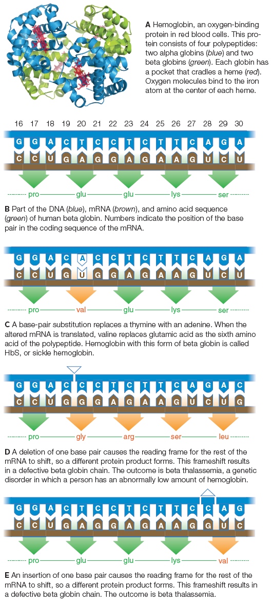
| Description: A Hemoglobin, an oxygen-binding protein in red blood cells. This pro-tein consists of four polypeptides: two alpha globins (blue) and two beta globins (green). Each globin has a pocket that cradles a heme (red). Oxygen molecules bind to the iron atom at the center of each heme. B Part of the DNA (blue), mRNA (brown), and amino acid sequence (green) of human beta globin. Numbers indicate the position of the base pair in the coding sequence of the mRNA. C A base-pair substitution replaces a thymine with an adenine. When the altered mRNA is translated, valine replaces glutamic acid as the sixth amino acid of the polypeptide. Hemoglobin with this form of beta globin is called HbS , or sickle hemoglobin. D A deletion of one base pair causes the reading frame for the rest of the mRNA to shift, so a different protein product forms. This frameshift results in a defective beta globin chain. The outcome is beta thalassemia, a genetic disorder in which a person has an abnormally low amount of hemoglobin. E An insertion of one base pair causes the reading frame for the rest of the mRNA to shift, so a different protein product forms. This frameshift results in a defective beta globin chain. The outcome is beta thalassemia. Picture Stats: Views: 94 Filesize: 334.61kB Height: 1174 Width: 532 Source: https://biology-forums.com/index.php?action=gallery;sa=view;id=49396 |