 |
| Previous Image | Next Image |
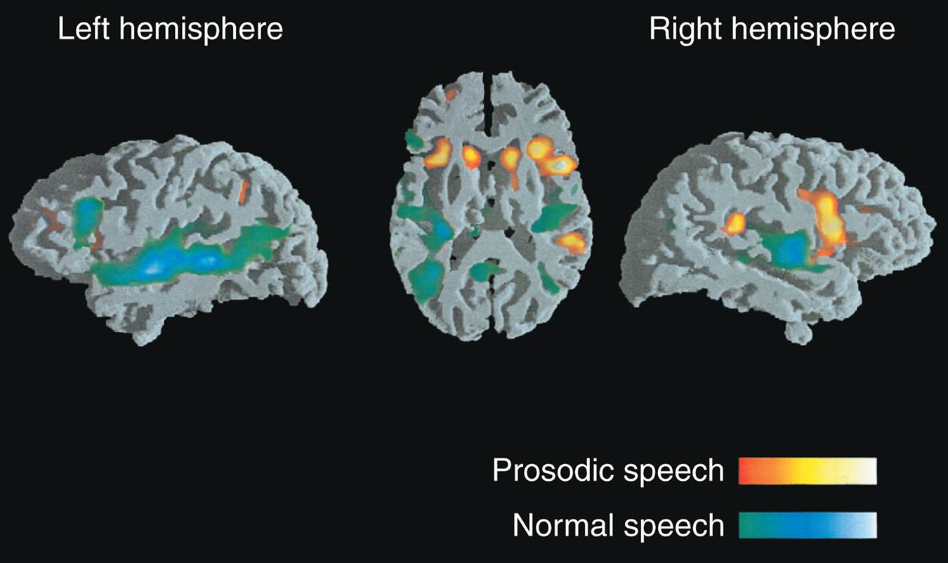
| Description: Functional MRI scans were made while subjects listened to normal speech (blue and green regions) or the prosodic elements of speech with the meaningful components filtered out (orange and yellow regions). (From Meyer, M., Alter, K., Friederici, A. D., Lohmann, G., and von Cramon, D. Y. Human Brain Mapping, 2002, 17, 73–88. Reprinted with permission.)
Picture Stats: Views: 726 Filesize: 73.71kB Height: 802 Width: 1350 Source: https://biology-forums.com/index.php?action=gallery;sa=view;id=21897 |