 |
| Previous Image | Next Image |
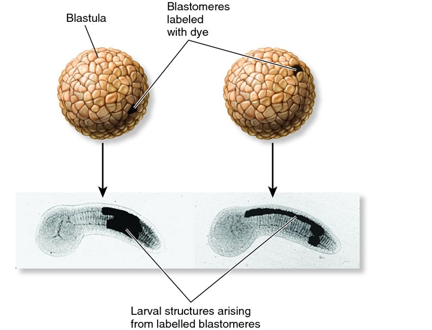
| Description: Fate mapping of single, labelled embryonic cells (blastomeres) shows that different cells in the embryo (in this case, a urochordate blastula) form different structures in the elongated larva, as shown by the dark regions. In the larva on the right, much of the dorsal nerve cord appears to be labelled.
Picture Stats: Views: 1138 Filesize: 56.24kB Height: 649 Width: 850 Source: https://biology-forums.com/index.php?action=gallery;sa=view;id=1312 |