 |
| Previous Image | Next Image |
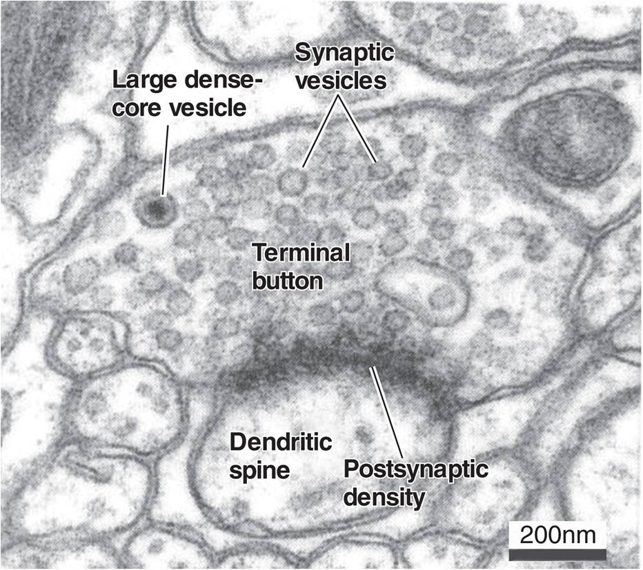
| Description: The photograph from an electron microscope shows a cross section of a synapse. The terminal button contains many synaptic vesicles, filled with the neurotransmitter, and a single large dense-core vesicle, filled with a peptide. (From De Camilli et al., in Synapses, edited by W. M. Cowan, T. C. Südhof, and C. F. Stevens. Baltimore, MD: Johns Hopkins University Press, 2001. Reprinted with permission.)
Picture Stats: Views: 566 Filesize: 134.04kB Height: 827 Width: 932 Source: https://biology-forums.com/index.php?action=gallery;sa=view;id=21034 |