 |
| Previous Image | Next Image |
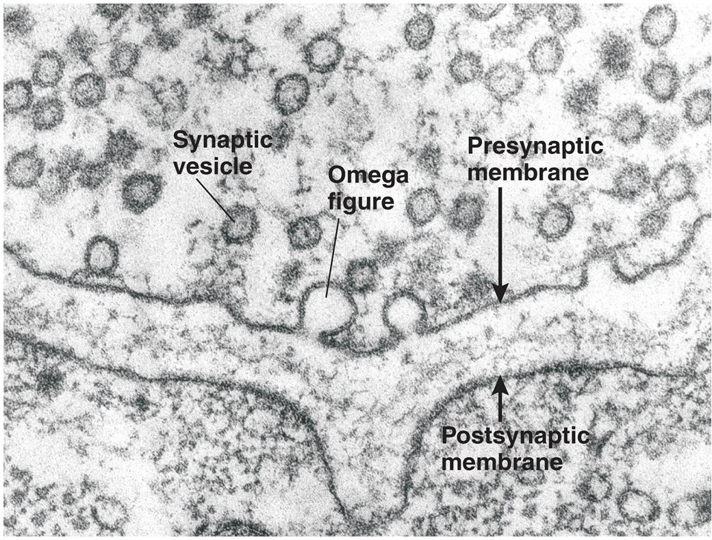
| Description: The photograph from an electron microscope shows a cross section of a synapse. The omega-shaped figures are synaptic vesicles fusing with the presynaptic membranes of terminal buttons that form synapses with frog muscle. (From Heuser, J. E., in Society for Neuroscience Symposia, Vol. II, edited by W. M. Cowan and J. A. Ferrendelli. Bethesda, MD: Society for Neuroscience, 1977. Reprinted with permission.)
Picture Stats: Views: 692 Filesize: 196.86kB Height: 789 Width: 1040 Source: https://biology-forums.com/index.php?action=gallery;sa=view;id=21035 |
