 |
| Previous Image | Next Image |
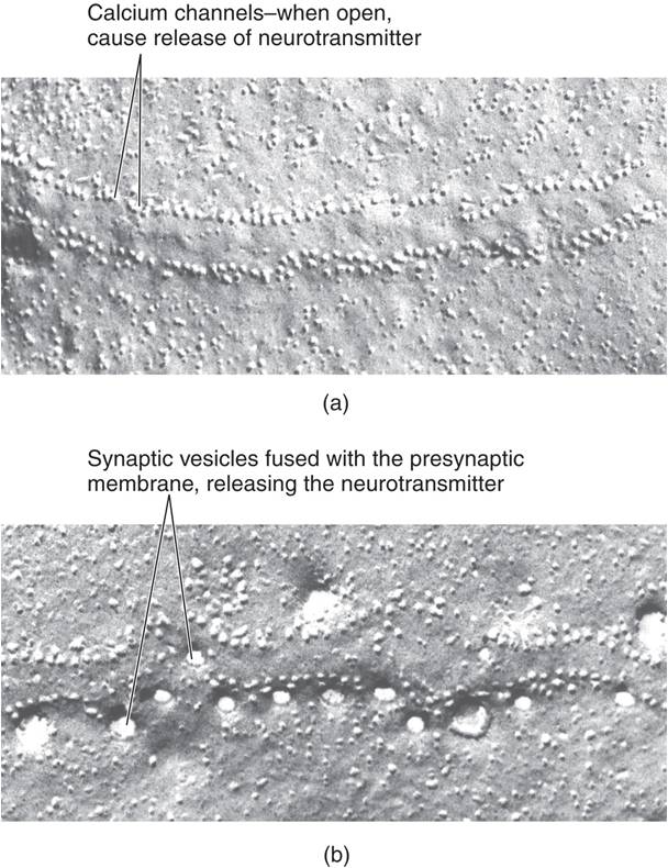
| Description: These photomicrographs show the release of neurotransmitter by a terminal button that forms a synapse with a frog muscle. The views are of the surface of the fusion zone of the terminal button. (a) Just before release. The two rows of dots are probably calcium channels. (b) During release. The larger circles are holes in the presynaptic membrane, revealing the contents of the synaptic vesicles that have fused with it. (From Heuser, J., and Reese, T. Journal of Cell Biology, 1981, 88, 564–580. Reprinted with permission.)
Picture Stats: Views: 629 Filesize: 89.36kB Height: 790 Width: 608 Source: https://biology-forums.com/index.php?action=gallery;sa=view;id=21037 |