 |
| Previous Image | Next Image |
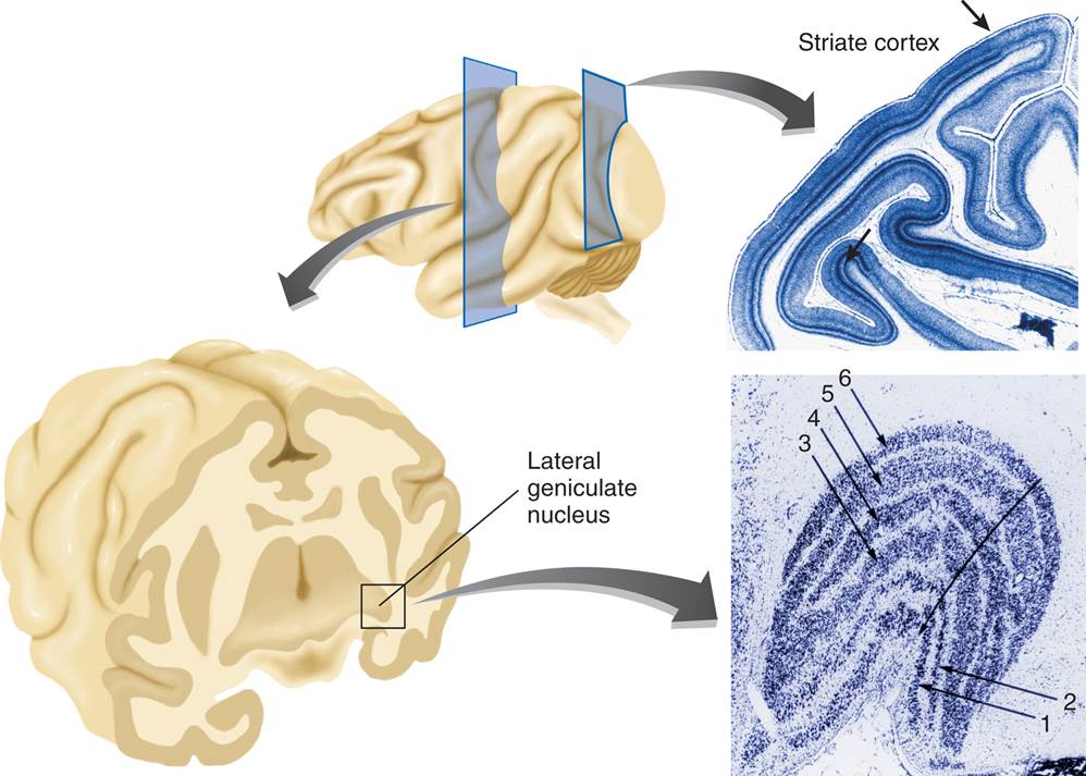
| Description: This photomicrograph shows a section through the right lateral geniculate nucleus of a rhesus monkey (cresyl violet stain). Layers 1, 4, and 6 receive input from the contralateral (left) eye, and layers 2, 3, and 5 receive input from the ipsilateral (right) eye. Layers 1 and 2 are the magnocellular layers; layers 3–6 are the parvocellular layers. The koniocellular sublayers are found ventral to each of the parvocellular and magnocellular layers. The receptive fields of all six principal layers are in almost perfect registration; cells located along the line of the unlabeled arrow have receptive fields centered on the same point. (Photomicrograph from Hubel, D. H., Wiesel, T. N., and Le Vay, S. Philosophical Transactions of the Royal Society of London, B, 1977, 278, 131–163. Reprinted with permission.)
Picture Stats: Views: 587 Filesize: 89.23kB Height: 713 Width: 998 Source: https://biology-forums.com/index.php?action=gallery;sa=view;id=21231 |