 |
| Previous Image | Next Image |
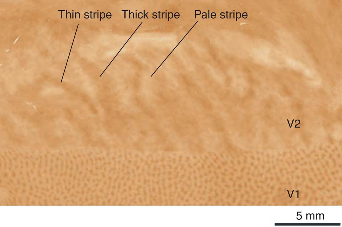
| Description: A photomicrograph (actually, a montage of several different tissue sections) showing a slice through the primary visual cortex (area V1) and a region of visual association cortex (V2) of a macaque monkey, stained for cytochrome oxidase. Area V1 shows spots (“blobs”), and area V2 shows three types of stripes: thick, thin (both dark), and pale. (From Sincich, L. C., and Horton, J. C. Annual Review of Neuroscience, Volume 28 © 2005, 303–326 by Annual Reviews www.annualreviews.o rg)
Picture Stats: Views: 589 Filesize: 49.96kB Height: 750 Width: 1130 Source: https://biology-forums.com/index.php?action=gallery;sa=view;id=21250 |