 |
| Previous Image | Next Image |
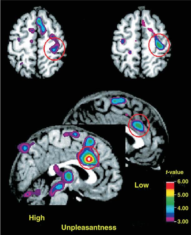
| Description: The PET scans show brain regions that respond to pain. Top: Dorsal views of the brain. Activation of the primary somatosensory cortex (circled in red) by a painful stimulus was not affected by a hypnotically suggested reduction in unpleasantness of a painful stimulus, indicating that this region responded to the sensory component of pain. Bottom: Midsagittal views of the brain. The anterior cingulate cortex (circled in red) showed much less activation when the unpleasantness of the painful stimulus was reduced by hypnotic suggestion. (From Rainville, P., Duncan, G. H., Price, D. D., Carrier, Benoit, and Bushnell, M. C. Science, 1997, 277, 968–971. Copyright © American Association for the Advancement of Science. Reprinted with permission.)
Picture Stats: Views: 601 Filesize: 55.05kB Height: 750 Width: 612 Source: https://biology-forums.com/index.php?action=gallery;sa=view;id=21403 |