 |
| Previous Image | Next Image |
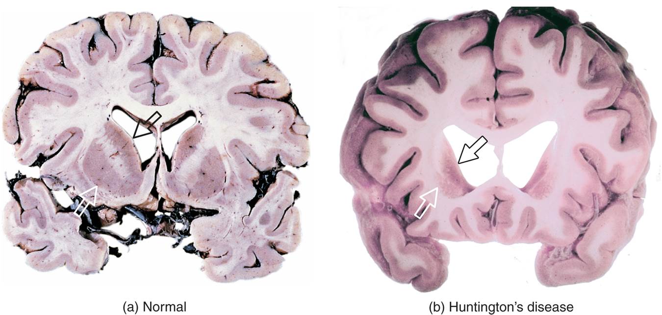
| Description: (a) A slice through a normal human brain, showing the normal appearance of the caudate nuclei and putamen (arrowheads) and lateral ventricles. (b) A slice through the brain of a person who had Huntington’s disease. The arrowheads indicate the location of the caudate nuclei and putamen, which are severely degenerated. As a consequence of the degeneration, the lateral ventricles (open spaces in the middle of the slice) have enlarged. (Courtesy of Harvard Medical School/Betty G. Martindale and Anthony D’Agostino, Good Samaritan Hospital, Portland, Oregon.)
Picture Stats: Views: 1497 Filesize: 93.56kB Height: 643 Width: 1350 Source: https://biology-forums.com/index.php?action=gallery;sa=view;id=21453 |