 |
| Previous Image | Next Image |
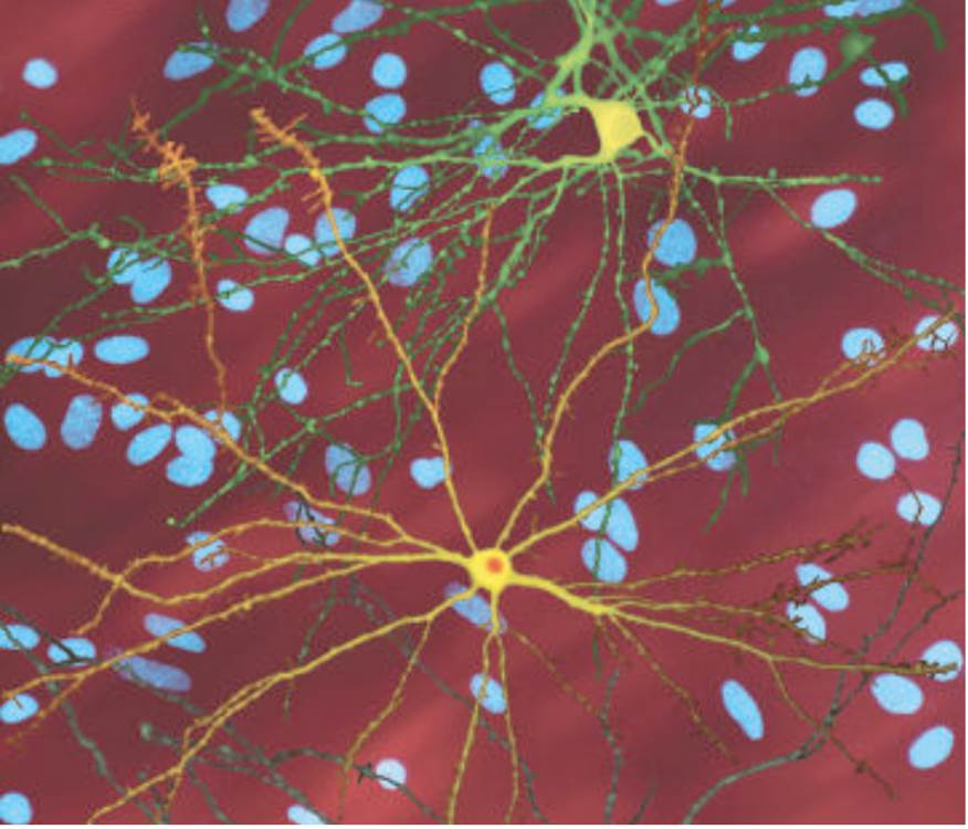
| Description: The photomicrograph shows two neurons that have been infected with genes that express fragments of abnormal huntingtin. The lower neuron shows an inclusion body (lorange), and the upper one does not. Arrasate et al. (2004) found that neurons with inclusion bodies survived longer than those without inclusion bodies. Blue ovals are the nuclei of uninfected neurons. (Photo courtesy of Steven Finkbeiner, Gladstone Institute of Neurological Disease and the University of California, San Francisco.)
Picture Stats: Views: 639 Filesize: 71.53kB Height: 750 Width: 876 Source: https://biology-forums.com/index.php?action=gallery;sa=view;id=21833 |