 |
| Previous Image | Next Image |
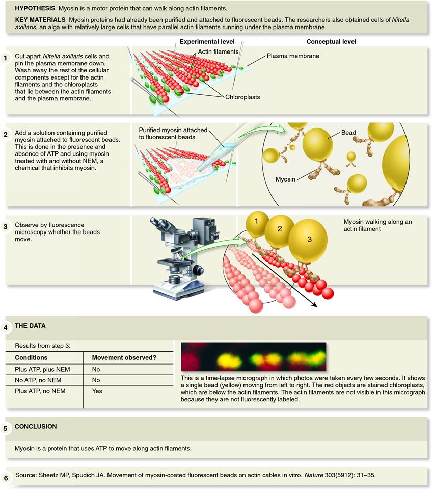
| Description: This figure illustrates the in vitro approach of Sheetz and Spudich. Their procedure involved cutting open the large Nitella cells and pinning down the plasma membrane to the substrate. They then carefully rinsed the cells. Except for chloroplasts, which are found between the plasma membrane and the actin filaments, the rest of the cellular contents were washed away, exposing the actin filaments. Next, a solution containing purified myosin attached to fluorescent beads was added and observed via fluorescence microscopy. Sheetz and Spudich conducted their experiments with and without ATP, because it was known that ATP was needed for muscle cell movement. In addition, the researchers tested the effects of N-ethylmaleimide (NEM), a chemical that was already known to bind to myosin and inhibit its function. The researchers looked for myosin movement under the various conditions. The results are summarized in part (4). Myosin was observed moving along actin filaments only when ATP was present and NEM was absent.
Picture Stats: Views: 2634 Filesize: 146.86kB Height: 965 Width: 850 Source: https://biology-forums.com/index.php?action=gallery;sa=view;id=240 Keywords: Movement of myosin-coated beads along actin filaments. |