 |
| Previous Image | Next Image |
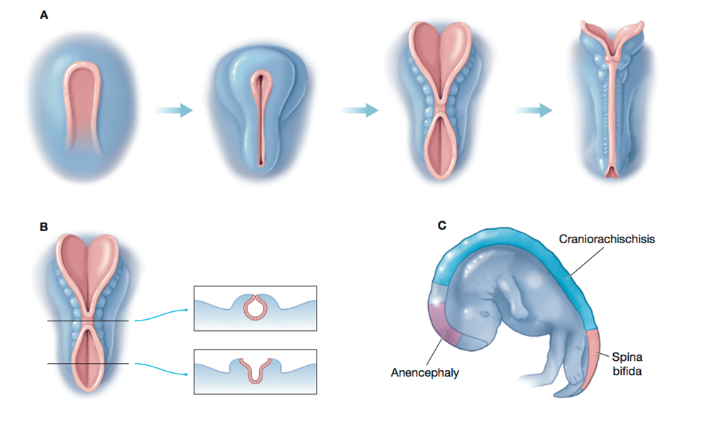
| Description: Row A shows the steps in neural tube formation and closure. Picture B shows points in development when the tube either closes (top) or fails to close (bottom). Picture C shows the regions where neural tube defects occur in an embryo at 4 weeks of age. Source: From Wallingford et al. (2013), p. 1049 Picture Stats: Views: 670 Filesize: 442.73kB Height: 598 Width: 999 Source: https://biology-forums.com/index.php?action=gallery;sa=view;id=24483 |