 |
| Previous Image | Next Image |
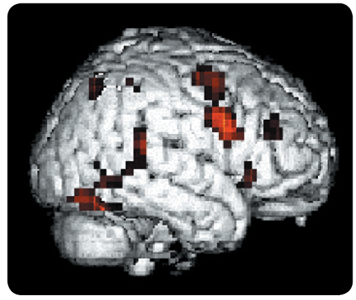
| Description: Functional magnetic resonance image (fMRI). This image illustrates the areas of cortex that became more active when the volunteers observed strings of letters and were asked to specify which strings were words; in the control condition, subjects viewed strings of asterisks (Kiehl et al., 1999). This fMRI illustrates surface activity; but images of sections through the brain can also be displayed. (Courtesy of Kent Kiehl and Peter Liddle, Department of Psychiatry, University of British Columbia.) Picture Stats: Views: 454 Filesize: 313.53kB Height: 436 Width: 517 Source: https://biology-forums.com/index.php?action=gallery;sa=view;id=30272 |
