 |
| Previous Image | Next Image |
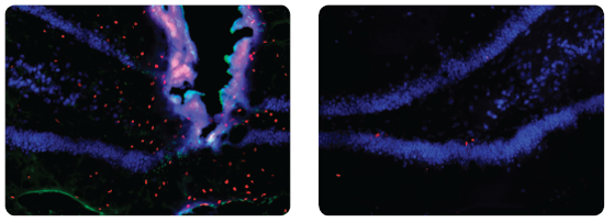
| Description: Increased neurogenesis in the dentate gyrus following damage. The left panel shows (1) an electrolytic lesion in the dentate gyrus (damaged neurons are stained turquoise) and (2) the resulting increase in the formation of new cells (stained red), many of which develop into mature neurons (stained dark blue). The right panel displays the comparable control area in the unlesioned hemisphere, showing the normal number of new cells (stained red). (These images are courtesy of my friends Carl Ernst and Brian Christie, Department of Psychology, University of British Columbia.) Picture Stats: Views: 424 Filesize: 177.01kB Height: 202 Width: 559 Source: https://biology-forums.com/index.php?action=gallery;sa=view;id=30386 |
