 |
| Previous Image | Next Image |
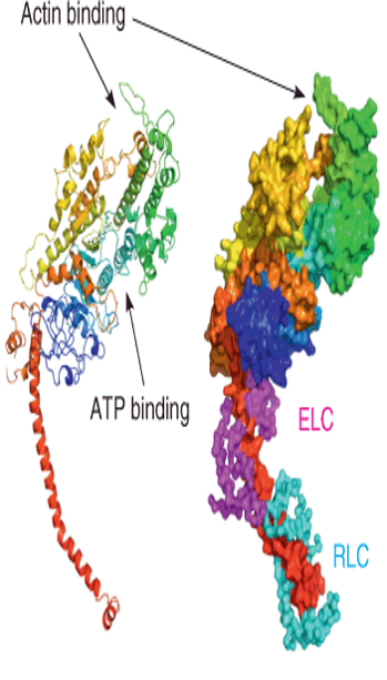
| Description: Left: a cartoon rendering of the heavy chain in rainbow coloring. The “neck” of the S1 fragment is shown by the extended red helix. Right: the heavy chain in a surface representation with the a–carbon backbone of the essential (ELC, magenta) and regulatory (RLC, cyan) light chains. The position of ATP binding is shown, as well as the point of contact with actin. Picture Stats: Views: 327 Filesize: 231.6kB Height: 613 Width: 349 Source: https://biology-forums.com/index.php?action=gallery;sa=view;id=34153 |