 |
| Previous Image | Next Image |
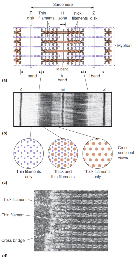
| Description: a) A model of the sarcomere. b) An electron micrograph showing the same features. c) A schematic drawing of cross sections of a sarcomere in the various regions shown in (a) and (b). Thick filaments are indicated by heavy brown dots, thin filaments by small purple dots. d) A higher magnification within an A band showing cross-bridges between actin and myosin filaments. Picture Stats: Views: 409 Filesize: 314.6kB Height: 863 Width: 448 Source: https://biology-forums.com/index.php?action=gallery;sa=view;id=34155 |