 |
| Previous Image | Next Image |
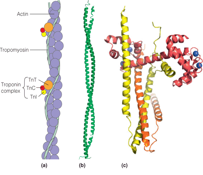
| Description: Panel (a): This schematic drawing shows the proteins present in the thin filaments of striated muscle: F-actin, tropomyosin, and troponins (Tn) I, C, and T. Panel (b): Crystal structure of a fragment of rat skeletal tropomyosin showing the coiled coil (PDB ID: 2B9C). Panel (C): The troponin complex from chicken skeletal muscle (PDB ID: 1YTZ). TnI is shown in yellow, TnT in orange and TnC in red with four bound Ca2+ ions shown as blue spheres. Picture Stats: Views: 274 Filesize: 271.41kB Height: 584 Width: 678 Source: https://biology-forums.com/index.php?action=gallery;sa=view;id=34158 |