 |
| Previous Image | Next Image |
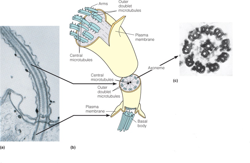
| Description: a) Electron micrograph of a longitudinal section of a cilium, showing microtubules running the length of the appendage. b) Schematic drawing of cilium structure. c) Electron micrograph of a cross section of the axoneme, showing the 9 + 2 arrangement of outer doublets and inner tubules. Picture Stats: Views: 224 Filesize: 290.77kB Height: 530 Width: 813 Source: https://biology-forums.com/index.php?action=gallery;sa=view;id=34164 |
