 |
| Previous Image | Next Image |
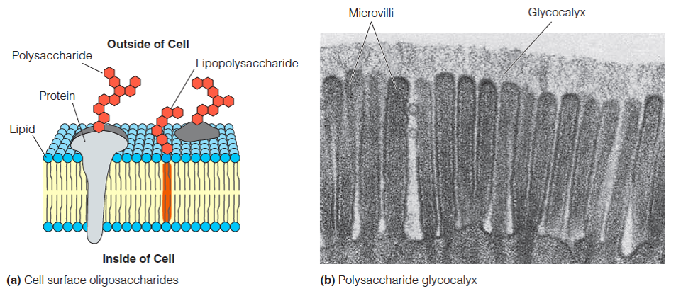
| Description: a. Schematic view of a lipid membrane. b. Electron micrograph of the surface of an intestinal epithelial cell. The cellular projections, called microvilli, are covered on their outer surface by a layer of branched polysaccharide chains attached to proteins in the cell membrane. This carbohydrate layer, called the glycocalyx, is found on many animal cell surfaces. Picture Stats: Views: 379 Filesize: 380.95kB Height: 411 Width: 953 Source: https://biology-forums.com/index.php?action=gallery;sa=view;id=34298 |
