 |
| Previous Image | Next Image |
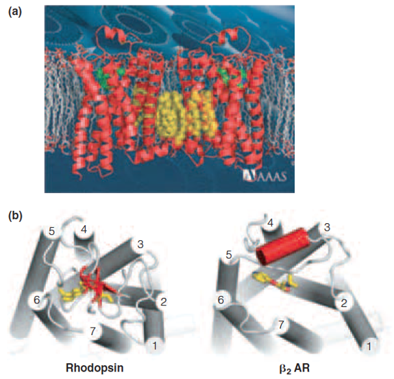
| Description: A model of two receptors embedded in the membrane and joined by cholesterol molecules (in yellow). The ligand carazolol is shown in green. Comparison of the top (extracellular) views of rhodopsin and the b2-adrenergic receptor (PDB ID 2RH1), showing similarities in arrangement of the transmembrane helices. Each ligand (11-cis-retinal in rhodopsin, carazolol in b2AR) is shown in red. A helical region of an extracellular domain, which helps to form the binding pocket for epinephrine, is shown in red. Picture Stats: Views: 352 Filesize: 541.39kB Height: 552 Width: 575 Source: https://biology-forums.com/index.php?action=gallery;sa=view;id=34870 |