 |
| Previous Image | Next Image |
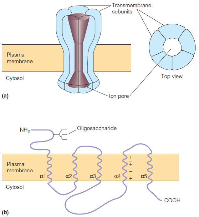
| Description: a. Schematic model of the receptor. Five subunits combine to form a transmembrane structure with an ion pore in the center. b. Structure of an individual subunit. There are four different kinds of subunits, but their sequences are all similar, and each individual subunit has the kind of structure depicted here. Five helixes (a1 to a5) in each subunit traverse the membrane. The charged residues on helix which tend to be on one surface, probably line the wall of the pore. Picture Stats: Views: 351 Filesize: 128.45kB Height: 695 Width: 636 Source: https://biology-forums.com/index.php?action=gallery;sa=view;id=34892 |
