 |
| Previous Image | Next Image |
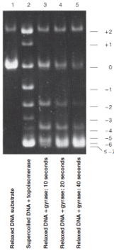
| Description: Lane 1 shows a relaxed circular DNA. Lane 2 shows the pattern from treatment of supercoiled DNA with type I topoisomerase. Lanes 3–5 show relaxed circles treated with DNA gyrase, a type II topoisomerase, for different lengths of time. Note that more different topoisomers can be seen in topoisomerase I reaction mixtures, as expected if changes in the linking number (L) occur in units of 1, whereas gyrase changes L in units of 2. Picture Stats: Views: 468 Filesize: 61.56kB Height: 354 Width: 161 Source: https://biology-forums.com/index.php?action=gallery;sa=view;id=34957 |