 |
| Previous Image | Next Image |
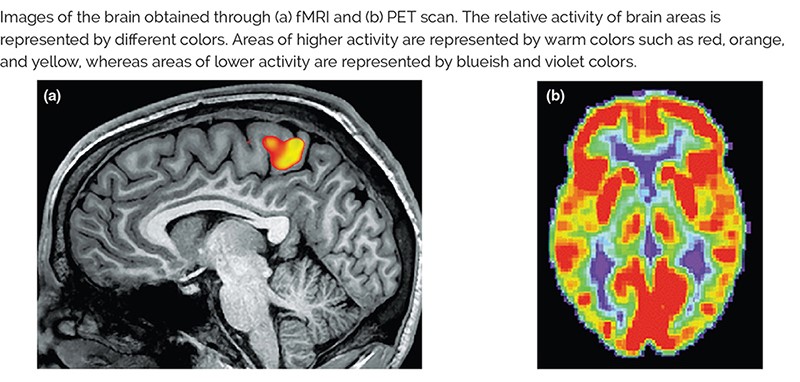
| Description: Images of the brarn obtamed through (a) WW and (bl PET scan The relatlve actlvlly or bram areas ls represented by ortrerent colors Areas of hlghel activity are represented by warm colors such as red, orange. and yellow. wneress areas of lower sctrvity are represented by bluelsh and vrolet colors
Picture Stats: Views: 141 Filesize: 97.48kB Height: 382 Width: 800 Source: https://biology-forums.com/index.php?action=gallery;sa=view;id=48357 |