 |
| Previous Image | Next Image |
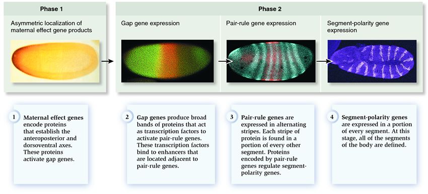
| Description: The micrographs depict the progression of Drosophila development during the first few hours following fertilization. The micrographs also show the expression of protein products of a maternal effect gene (step 1) or segmentation genes (steps 2–4). In step 1, the protein is stained brown and is found in the left side of the early embryo. In step 2, one protein encoded by a gap gene is stained in green and another is stained in red. The yellow region is the point at which the two different gap proteins overlap. In step (3), a protein encoded by a pair-rule gene is stained in light blue. In step (4), a protein encoded by a segment-polarity gene is stained pink. When comparing steps (3) and (4), note that the embryo has made a 180° turn, folding back on itself.
Picture Stats: Views: 6473 Filesize: 66.02kB Height: 386 Width: 850 Source: https://biology-forums.com/index.php?action=gallery;sa=view;id=573 Keywords: Overview of segmentation in Drosophila |