 |
| Previous Image | Next Image |
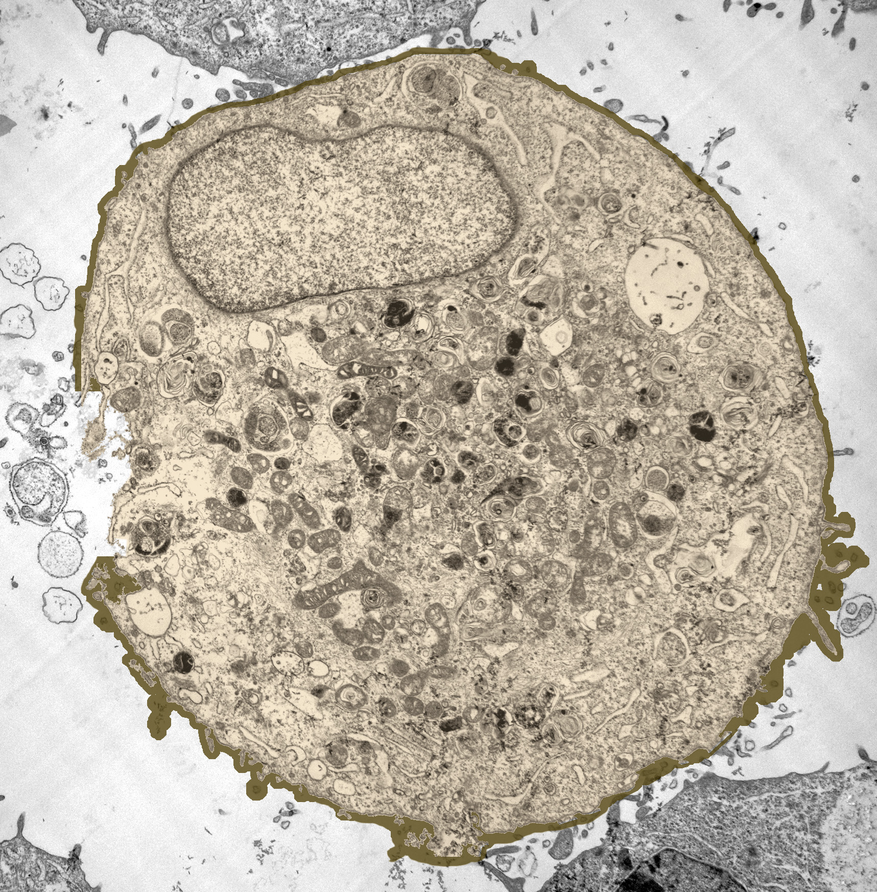
| Description: Transmission electron micrograph showing a prostate cancer cell immediately after exposure to ultrasound. The image has been color enhanced to show the spot where the cell membrane has been removed.
Picture Stats: Views: 2510 Filesize: 1.28MB Height: 1746 Width: 1717 Source: https://biology-forums.com/index.php?action=gallery;sa=view;id=8372 Keywords: Prostate Cancer Cell |
