 |
| Previous Image | Next Image |
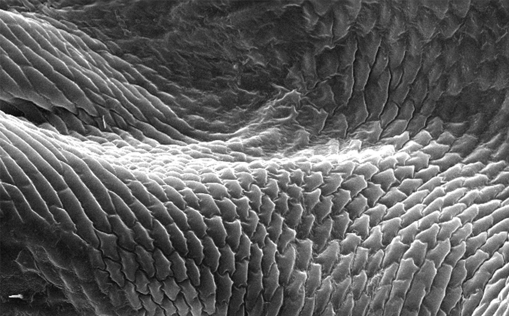
| Description: Magnified 598x, this scanning electron micrograph depicts an enlarged view of the chitinous, exoskeletal surface of a male louse, Pediculus humanus var. corporis. In this particular view, the exoskeleton appears to be composed of interlocking plates.
Picture Stats: Views: 1129 Filesize: 195.17kB Height: 616 Width: 990 Source: https://biology-forums.com/index.php?action=gallery;sa=view;id=8384 Keywords: chitinous exoskeletal surface of a male louse |
