Answer to Question 1
ANS: A
A. Correct response: The fact that the capnogram tracing abruptly spiked from 0 exhaled CO2 to around 3 CO2 indicates that the endotracheal tube has entered the patient's trachea. Actually, with adequate perfusion and ventilation, one would expect to see an exhaled CO2 percent between 4.5 to 5.0.
B. Incorrect response: If the endotracheal tube was placed into the esophagus, the capnogram would continue to read essentially 0 exhaled CO2. The capnogram would effectively demonstrate a flat line superimposed on the baseline. The first half of the drawing indicates esophageal intubation.
C. Incorrect response: If the therapist had intubated the right mainstem bronchus instead of the trachea, the capnogram would still indicate the tracing shown with this question because CO2 from the right lung would be detected by the capnometer. A portable AP chest x-ray must be performed to confirm the location of the tip of the endotracheal tube.
D. Incorrect response: See explanation A.
Answer to Question 2
ANS: C
A. Incorrect response: See explanation C.
B. Incorrect response: See explanation C.
C. Correct response: The initial downward deflection on the flow-time tracing indicates the volume of gas compressed in the ventilator circuit at the end of inspiration. When inspiration ends, the compressed volume exits the ventilator breathing circuit rapidly. In fact, this volume is expelled more rapidly from the circuit than the patient's own exhaled volume. This sudden flow of gas leaving the breathing circuit causes an initial steep decline in along the flow tracing, followed by a gradual tapering of flow to zero.
D. Incorrect response: See explanation C.

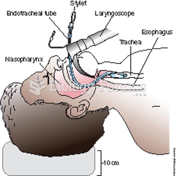 Placing an endotracheal tube in the trachea with direct visualization by laryngoscopy.
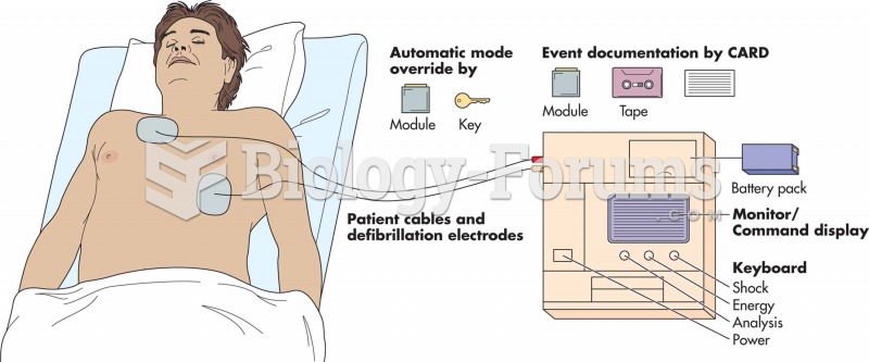 Schematic of an automated external defibrillator (AED) attached to a patient.
 The fallopian tube
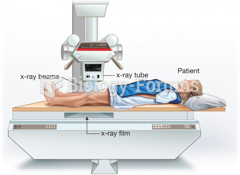 The patient is placed between the X-ray tube, which emits a cone-shaped X-ray beam, and the film or ...
 Wintrobe tube and Westergren tests are used to measure the ESR.
 I need help fill up the chart attached phtoto long question