Answer to Question 1
Post mortem analyses are typically performed on individuals who have brain lesions as a result of injury, disease or surgery or who have abnormal brain structures as a result of congenital differences. Although some such lesions can be created in animals for the purposes of research, they cannot ethically be created in humans for the purpose of research. Neuroimaging techniques, while they are continuously advancing in their ability to provide both static and dynamic images of the brain, may miss subtle structural differences that are visual when stained brain slices are viewed under the microscope.
Answer to Question 2
The frontal lobe, toward the front of the brain, is associated with motor processing and higher thought processes, such as abstract reasoning, problem solving, planning, and judgment. It tends to be involved when sequences of thoughts or actions are called for. It is critical in producing speech. The prefrontal cortex, the region toward the front of the frontal lobe, is involved in complex motor control and tasks that require integration of information over time.
The parietal lobe, at the upper back portion of the brain, is associated with somatosensory processing. The primary somatosensory cortex receives information from the senses about pressure, texture, temperature, and pain. It is located right behind the frontal lobe's primary motor cortex. If your somatosensory cortex were electrically stimulated, you probably would report feeling as if you had been touched. The parietal lobe also helps you perceive space and your relationship to ithow you are situated relative to the space you are occupying. It is also involved in consciousness and paying attention.
The temporal lobe is located below the parietal lobe, directly under your temples. It is associated with auditory processing and comprehending language. Some parts are more sensitive to sounds of higher pitch, others to sounds of lower pitch. The auditory region is primarily contralateral. Both sides of the auditory area have at least some representation from each ear. If your auditory cortex were stimulated electrically, you would report having heard some sort of sound. The temporal lobe is also involved in retaining visual memories. For example, if you are trying to keep in memory
The occipital lobe is associated with visual processing. The occipital lobe contains numerous visual areas, each specialized to analyze specific aspects of a scene, including color, motion, location, and form. Projection areas are the areas in the lobes in which sensory processing occurs. These areas are referred to as projection areas because the nerves contain sensory information going to (projecting to) the thalamus. It is from here that the sensory information is communicated to the appropriate area in the relevant lobe. Similarly, the projection areas communicate motor information downward through the spinal cord to the appropriate muscles via the peripheral nervous system (PNS). The visual cortex is primarily in the occipital lobe. Some neural fibers carrying visual information travel ipsilaterally from the left eye to the left cerebral hemisphere and from the right eye to the right cerebral hemisphere. Other fibers cross over the optic chiasma and go contralaterally to the opposite hemisphere. In particular, neural fibers go from the left side of the visual field for each eye to the right side of the visual cortex. Complementarily, the nerves from the right side of each eye's visual field send information to the left side of the visual cortex.

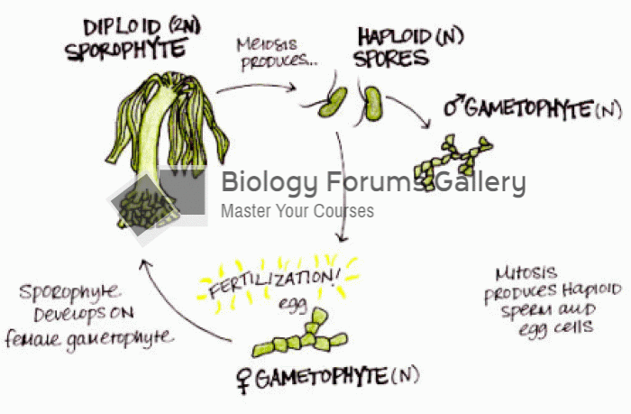 Plants: alternation of generations
 Community colleges have opened higher education to millions of students who would not otherwise have ...
 Generations of Fluoroquinolones
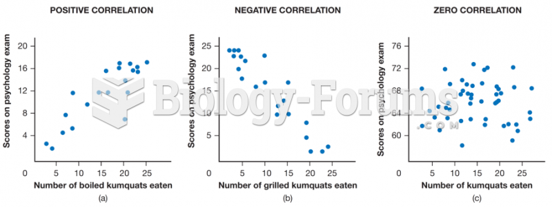 Correlations between scores on a psychology test and number of boiled kumquats eaten per month
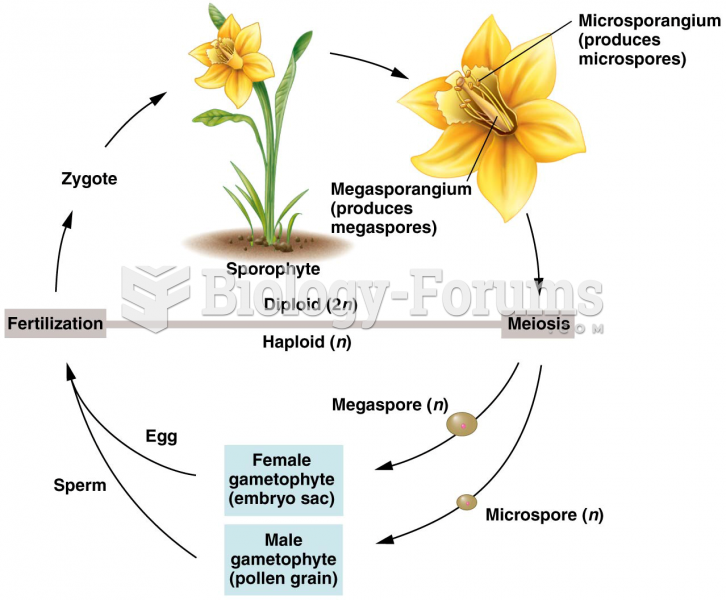 Alternation of generations between the diploid sporophyte
 The total weight of Peter, David and Henry is 123 kg, Peter is 15 kg heavier than David. David ...