Positioning the femur at a 60- to 70-degree angle with the imaging table for the PA axial knee projection (Holmblad method)
1. superimposes the proximal surfaces of the intercondylar fossa.
2. places the patellar apex superior to the intercondylar fossa.
3. superimposes the lateral and the medial surfaces of the intercondylar fossa.
4. superimposes the anterior and posterior margins of the tibia plateau.
a. 1 only
b. 1 and 2 only
c. 3 and 4 only
d. 1, 2, and 3 only
Question 2
A lateral knee projection obtained with the patella positioned too close to the IR (leg externally rotated) will demonstrate the
1. fibula with increased tibial superimposition.
2. fibula with decreased tibial superimposition.
3. medial femoral condyle anterior to the lateral femoral condyle.
4. medial condyle distal to the lateral femoral condyle.
a. 1 and 3 only
b. 1 and 4 only
c. 2 and 3 only
d. 2 and 4 only

 Redrape the back, face the head of the table, and apply passive touch as finishing technique. Gently ...
 Finish with bilateral stroking to the entire back. Stand at head of the table with fingers pointing ...
 With the arm placed on the table, apply effleurage over the entire upper limb as a reconnecting and ...
 Deep effleurage with forearm to hamstring muscles. Apply from above knee to superior attachments.
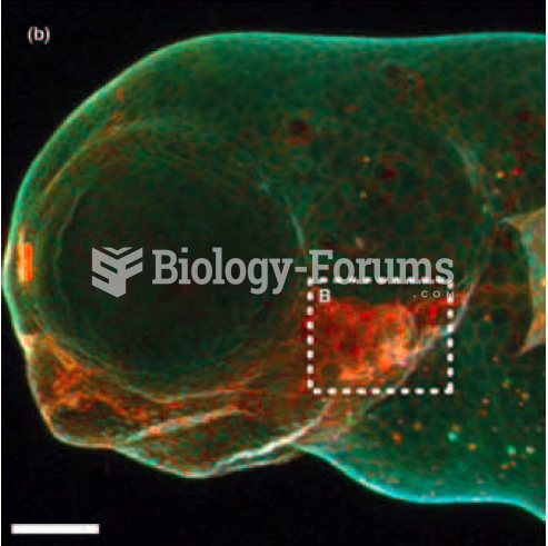 Imaging cell surface glycoproteins in living cells
 Seven bacteriophage deletion mutations (1 to 7 in the table below) are tested for their ability ...