This topic contains a solution. Click here to go to the answer
|
|
|

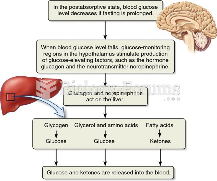 The role of the liver in fuel supply to the blood.
 Electron Microscope Images of a Diatom
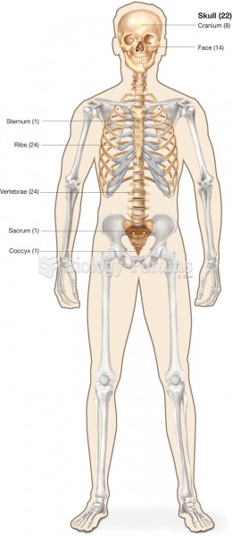 Bones of the axial skeleton.
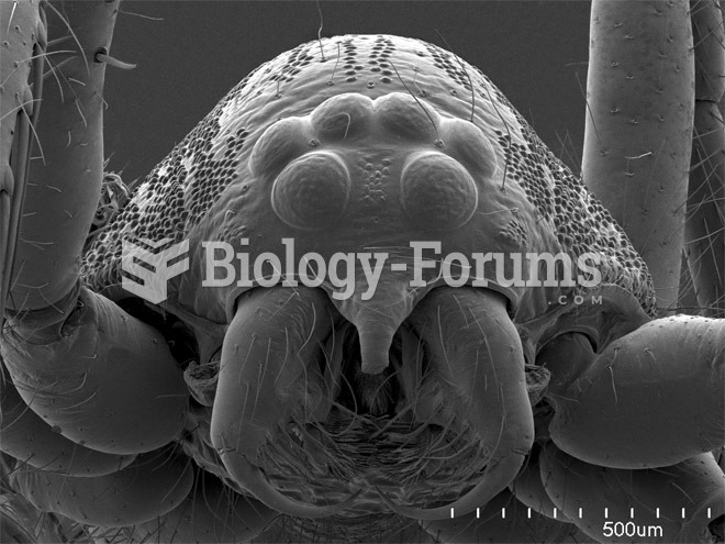 Scorpions, Spiders and Sharks: Electron-Microscope Images
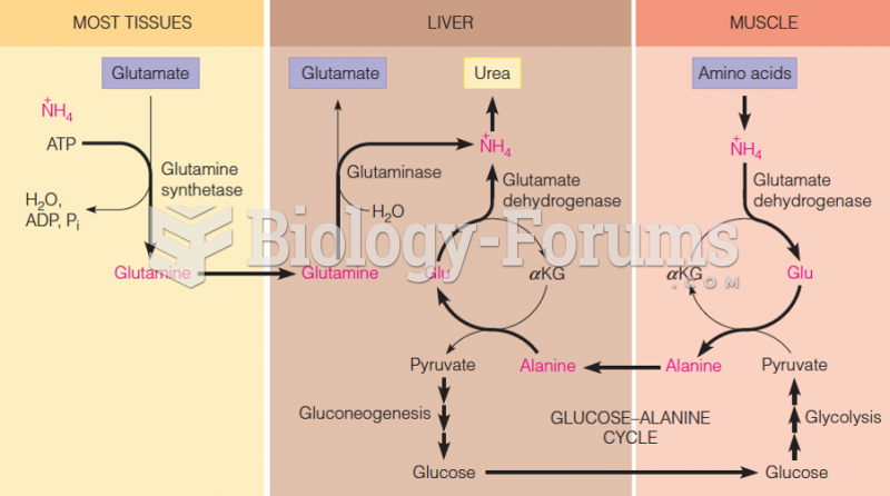 Transport of ammonia to the liver for urea synthesis