|
|
|

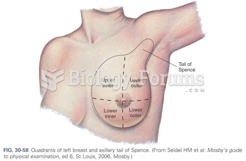 Quadrants of left breast and axillary tail of spence
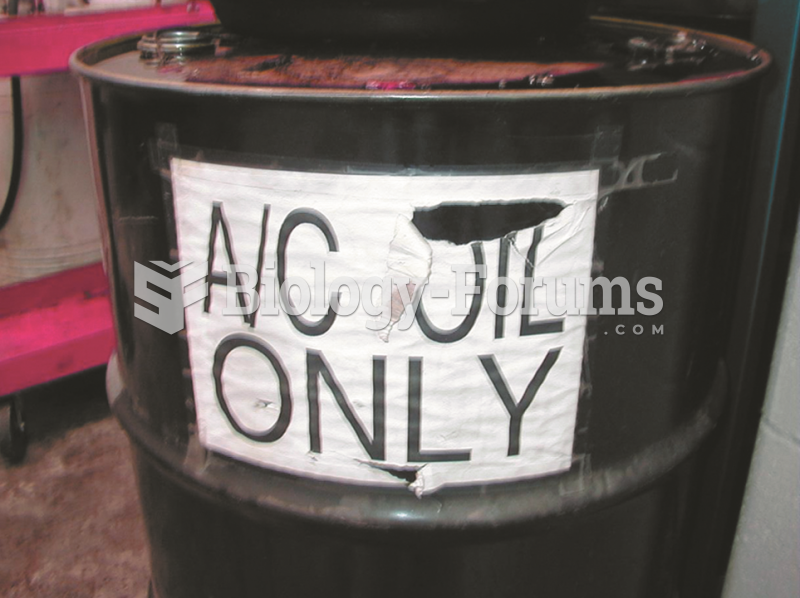 Air-conditioning refrigerant oil must be kept separated from other oils because it contains traces ...
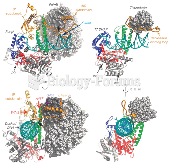 Structures of human DNA polymerase g (left) and T7 phage DNA polymerase (right) holoenzymes
 Left wing right wing
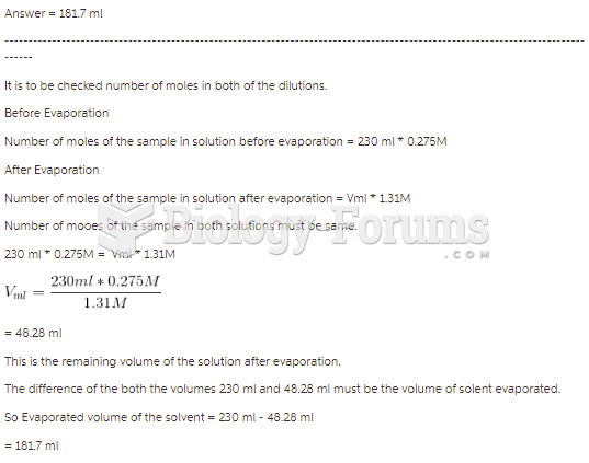 A 230.0-mL sample of a 0.275 M solution is left on a hot plate overnight; the following morning ...
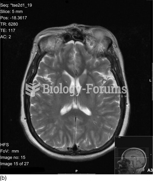 Axial M R I of the brain