|
|
|

 Forest fragments left by clear-cutting forest from the surrounding landscape have very different phy
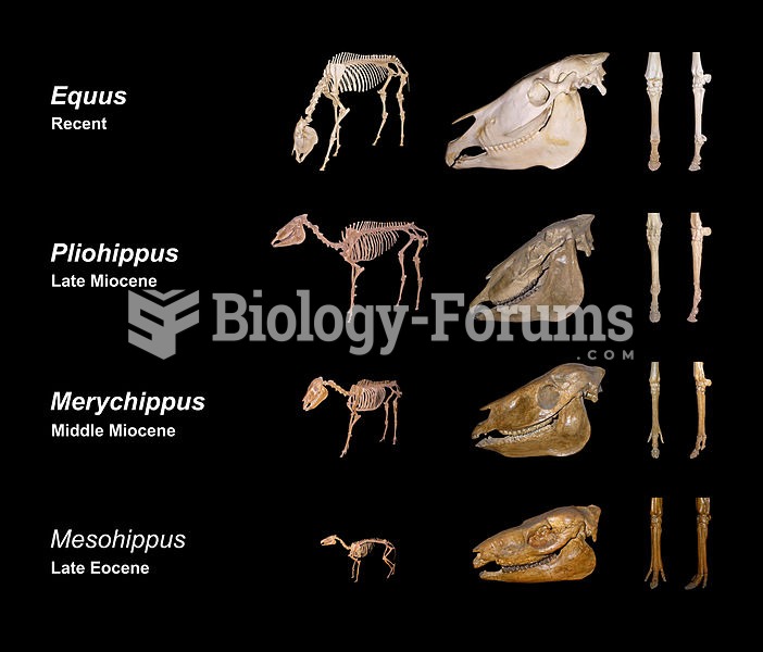 From left to right: Size development, biometrical changes in the cranium, reduction of toes ...
 Effects of Therapy for Stuttering A functional MRI scan shows regions of the superior temporal lobe
 Liver
Liver
 Liver
Liver
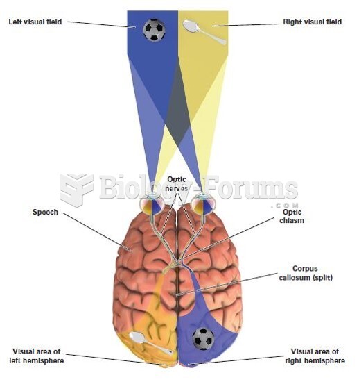 Right and Left Visual Fields