There have been lots of different things, most of the earliest ideas came from looking at the brains of people who had been injured, knowing that there were certain things regarding personality or intelligence that they could no longer do. Lots of animals were used in experiments, purposefully given brain damage to see how this affected their behaviour and abilities.
In recent decades there have been some inventions which have given a much better understanding of how human brains function. These are electronic maps called CT (computer tomography) and PET (positron emission tomography). CT scans (called "cat" scans) are kind of like an x-ray of the brain, but PET scans give a 3D picture and can be used while the patient is awake to show brain activity in response to stimuli. For example, they'll ask the person to think of an apple, and watch what parts of the brain are active, then they'll get them to click their fingers, then think about clicking their fingers, and compare how the brain activity differs between clicking fingers or just thinking about clicking them

 Much Exploration for Oil and Gas Takes Place in Remote Parts of the World
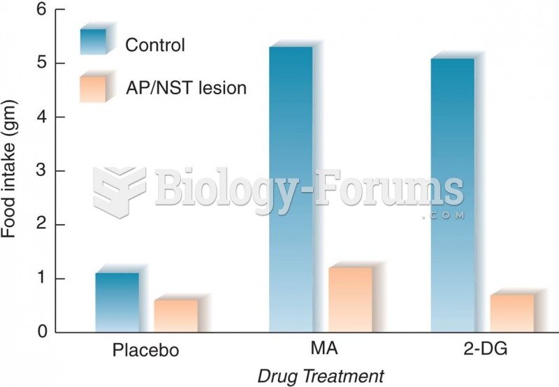 Role of the Brain Stem in Hunger
 Children in Baltimore (1909) pull the stringy parts from beans in preparation for canning. Photograp
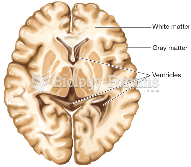 Gray and White Matter of the Brain
 Growth of the body and brain is dramatic over the first 2 years.
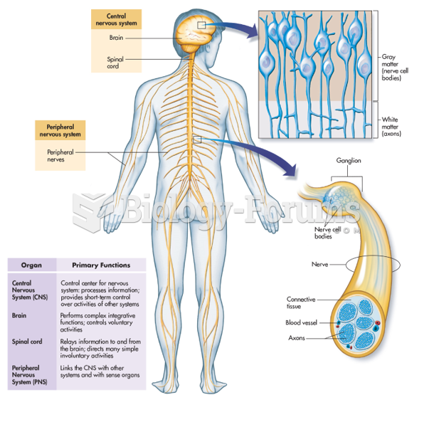 The Central Nervous System Includes the Brain and Spinal Cord