The microscopic examination of a Wright-stained blood smear revealed bright red erythrocytes and pale leukocyte nuclei. What is the best explanation for this appearance?
a. The buffer is too acidic.
b. The staining process was prolonged.
c. The blood smear was too thick.
d. The Wright stain was too alkaline.
Question 2
Which of the following receptacles is most appropriate when disposing of used needles?
a. A regular garbage can
b. A biohazard bag
c. A biohazard sharps container
d. A regular garbage bag

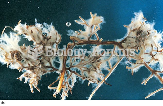 Mutualistic associations between fungi and plant roots: (a) arbuscular mycorrhizal fungus stained so
 Alapaha Blue Blood Bulldog
 Taking a Patient's Blood Pressure
 Breast self-examination.
 Testing for Occult Blood
 The primary factors affecting blood pressure.