Answer to Question 1
Answer:
1. Trauma occurs.
2. Subendothelial collagen is exposed.
3. Vasoconstriction is initiated to minimize blood loss.
4. Platelets become activated as a result of the subendothelial collagen (a) flat, discoid shape spiked appearance, (b) actin and myosin go from a random arrangement to a parallel arrangement; (c) tubulin forms a circumferential band around the organelle zone to assist in expelling granular contents to the surface of the platelets.
5. Activated platelets adhere to subendothelial collagen with the help of VWF and GP Ib/IX.
6. Once adhesion takes place, more platelets are recruited to the site, and aggregation is initiated. Substances needed for platelet aggregation is fibrinogen and GP IIb/IIIa.
7. Platelets secrete their granular contents and initiate secondary hemostasis.
8. Secondary hemostasis is activated by contact activation and subsequent progression into the coagulation cascade via the intrinsic and extrinsic pathways.
9. The end result is the formation of an insoluble fibrin polymer that stabilizes the platelet plug.
10. Fibroblasts grow and regenerate with the help of PDGF and other tissue repair chemokines.
11. Once repair is completed, the fibrinolysis pathway is activated.
12. Fibrin and tPA induce activation of plasmin from plasminogen.
13. Plasmin chemically digests the fibrin polymer into measurable fragments that can be detected in vitro.
14. Normal homeostasis is restored.
Answer to Question 2
Answer: Tunica adventitia: thick outer layer of the blood vessels responsible for maintaining blood pressure; is thicker in veins than in arteries
Tunica media: middle layer responsible for vasoconstriction and vasodilation; is thicker in arteries than in veins
Tunica intima: has direct interaction with peripheral blood constituents; all three types of vessels (arteries, veins, and capillaries) in close tunica intima layers

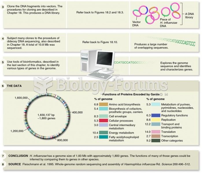 Determination of the complete genome sequence of Haemophilus influenzae by Venter, Smith, and collea
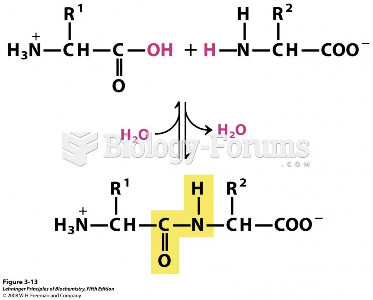 3-13 Formation of a peptide bond by condensation
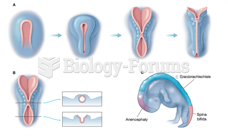 Formation of the Neural Tube and Neural Tube Defects
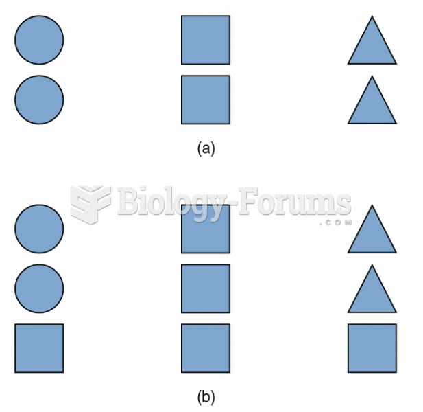 Concept Formation
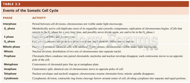 Events of the Somatic Cell Cycle
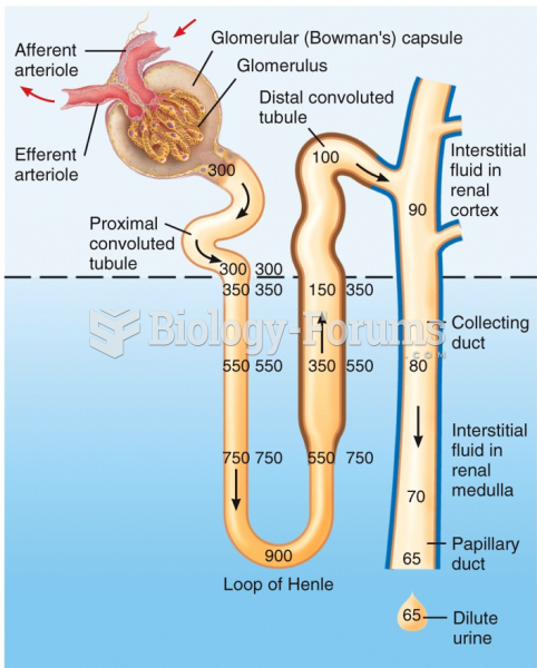 Formation of Dilute Urine