Answer to Question 1
The motor cortex can be mapped to show where, and in what proportions, different parts of the body are represented in the brain. Maps of this kind are called homunculi (homunculus is Latin for little person) because they depict the body parts of a person mapped on the brain. For both motor and sensory function, the size of the body part in the homunculus is related to function. Those areas of the body that are particularly sensitive or that are used in fine, complex motions have larger representations in the brain.
Answer to Question 2
Metabolic imaging techniques rely on changes that take place within the brain as a result of increased consumption of glucose and oxygen in active areas of the brain. The basic idea is that active areas in the brain consume more glucose and oxygen than do inactive areas during some tasks. An area specifically required by one task ought to be more active during that task than during more generalized processing and thus should require more glucose and oxygen.
PET scans measure increases in oxygen consumption in active brain areas during particular kinds of information. To track their use of oxygen, participants are given a mildly radioactive form of oxygen that emits positrons as it is metabolized (positrons are particles that have roughly the same size and mass as electrons, but that are positively rather than negatively charged). Next, the brain is scanned to detect positrons. A computer analyzes the data to produce images of the physiological functioning of the brain in action. PET scans are not highly precise because they require a minimum of about half a minute to produce data regarding glucose consumption. If an area of the brain shows different amounts of activity over the course of time measurement, the activity levels are averaged, potentially leading to conclusions that are less than precise.
Functional magnetic resonance imaging (fMRI) is a neuroimaging technique that uses magnetic fields to construct a detailed representation in three dimensions of levels of activity in various parts of the brain at a given moment in time. This technique builds on MRI, but it uses increases in oxygen consumption to construct images of brain activity. The basic idea is the same as in PET scans, but the fMRI technique does not require the use of radioactive particles. Rather, the participant performs a task while placed inside an MRI machine. This machine typically looks like a tunnel. When someone is wholly or partially inserted in the tunnel, he or she is surrounded by a doughnut-shaped magnet. An fMRI creates a magnetic field that induces changes in the particles of oxygen atoms. More active areas draw more oxygenated blood than do less active areas in the brain. So shortly after a brain area has been active, a reduced amount of oxygen should be detectable in this area. This observation forms the basis for fMRI measurements. These measurements then are computer analyzed to provide the most precise information currently available about the physiological functioning of the brain's activity during task performance.
A related procedure is pharmacological MRI (phMRI). The phMRI combines fMRI methods with the study of psychopharmacologic al agents. These studies examine the influence and role of particular psychopharmacologic al agents on the brain. phMRIs have been used to examine the role of agonists (which strengthen responses) and antagonists (which weaken responses) on the same receptor cells. These studies have allowed for the examination of drugs used for treatment. The investigators can predict the responses of patients to neurochemical treatments through examination of the person's brain makeup. Overall, these methods aid in the understanding of brain areas and the effects of psychopharmacologic al agents on brain functioning.
Another procedure related to fMRI is diffusion tensor imaging (DTI). DTI examines the restricted dispersion of water in tissue and, of special interest, in axons. Water in the brain cannot move freely, but rather, its movement is restricted by the axons and their myelin sheaths. DTI measures how far protons have moved in a particular direction within a specific time interval. This technique has been useful in the mapping of the white matter of the brain and in examining neural circuits. Some applications of this technique include examination of traumatic brain injury, schizophrenia, brain maturation, and multiple sclerosis
A recently developed technique for studying brain activity bypasses some of the problems with other techniques. Transcranial magnetic stimulation (TMS) temporarily disrupts the normal activity of the brain in a limited area. Therefore, it can imitate lesions in the brain or stimulate brain regions. TMS requires placing a coil on a person's head and then allowing an electrical current to pass through it. The current generates a magnetic field. This field disrupts the small area (usually no more than a cubic centimeter) beneath it. The researcher can then look at cognitive functioning when the particular area is disrupted.
Magnetoencephalogra phy (MEG) measures brain activity from outside the head (similar to EEG) by picking up magnetic fields emitted by changes in brain activity. This technique allows localization of brain signals so that it is possible to know what different parts of the brain are doing at different times. It is one of the most precise of the measuring methods. MEG is used to help surgeons locate pathological structures in the brain.

 Size varies greatly among horse breeds, as with this full-sized horse and a miniature horse.
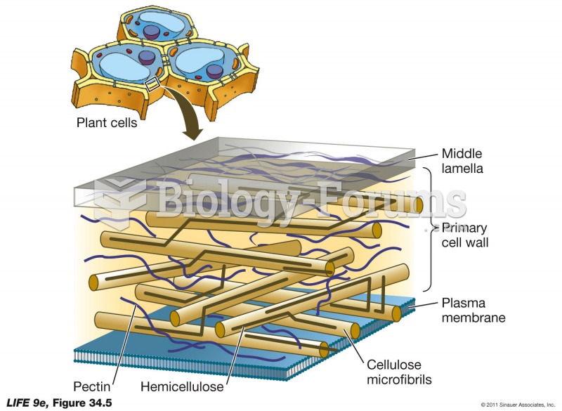 Plant Cell Wall Structure
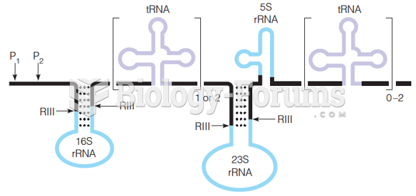 Structure of E. coli 30S pre-rRNA
 A Structure/Function Label Claim
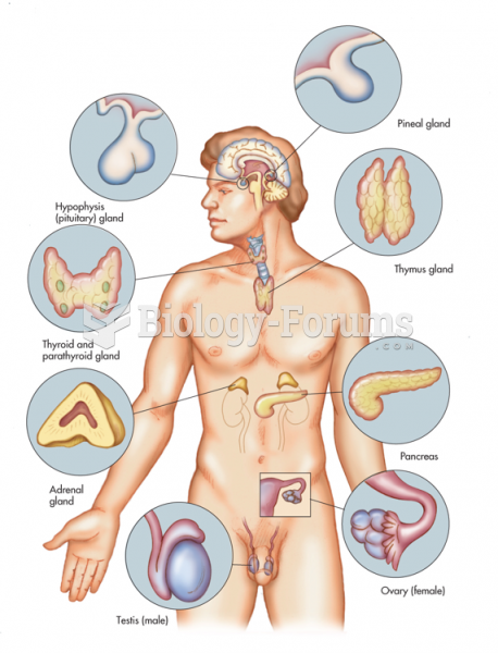 Primary Glands of the Endocrine System
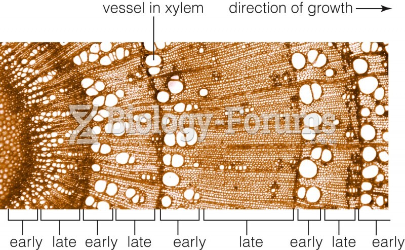 Structure of a woody stem