|
|
|

 Basal cell carcinoma, a frequent type of skin cancer that rarely metastasizes.
 Music while studying
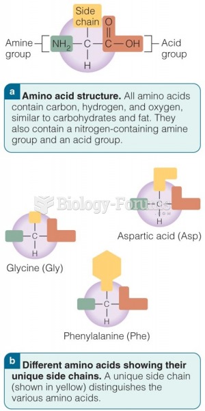 The Structure of an Amino Acid
 High stress, by household type (age-standardized), age 18+, Canada, 1994-1995
 What type of online info about a job candidate should employment mgmt consider when screening
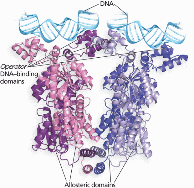 The homotetrameric structure of the lac repressor protein