This topic contains a solution. Click here to go to the answer
|
|
|

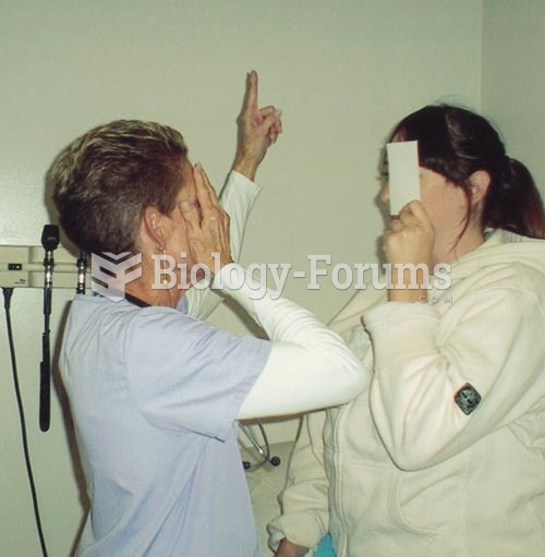 Testing Visual Fields by Confrontation: The nurse and patient should be approximately at an eye to e
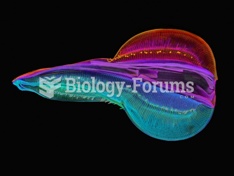 Electron Microscope Images of a Diatom
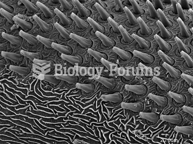 Scorpions, Spiders and Sharks: Electron-Microscope Images
 Relation between form, particular objects, and images in Plato’s Line.
 What level of barometric pressure effects body?
 The tertiary level of protein structure for the respiratory pigment myoglobin