A poorly positioned AP knee projection demonstrating a larger lateral femoral condyle than me-dial condyle
a. was obtained with the patient's leg externally rotated.
b. may also demonstrate the fibular head without tibial superimposition.
c. will also demonstrate a closed knee joint.
d. will also demonstrate the fibular head 1 inch (2.5 cm) distal to the tibial plateau.
Question 2
A poorly positioned 45-degree AP oblique ankle projection demonstrates the calcaneus obscuring the distal aspect of the lateral mortise and the distal fibula. How should the positioning setup be adjusted to obtain an optimal projection?
a. Increase the degree of internal leg rotation.
b. Decrease the degree of internal leg rotation.
c. Dorsiflex the foot to a 90-degree angle with the lower leg.
d. Center the central ray more distally on the ankle.

 Cardinal Fields of Gaze, Right Lateral Gaze
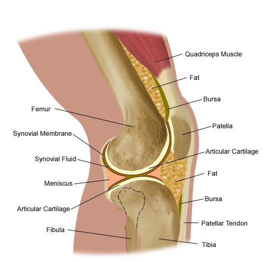 Knee
Knee
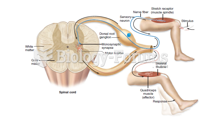 The knee-jerk reflex
 Ophthalmoscope demonstrating aperture
 Client positioned and draped
 In F- F+ mating, demonstrating how the recipient F- cell is converted to F+