A less than optimal axial calcaneus projection demonstrates an obscured talocalcaneal joint space and an elongated calcaneus tuberosity. The projection was obtained with the
a. patient's foot dorsiflexed beyond the required vertical position.
b. central ray angled less than the routinely required amount.
c. patient's foot in plantar flexion.
d. leg and ankle medially rotated.
Question 2
An accurately positioned AP knee projection demonstrates all the following except the
a. medial and lateral femoral epicondyles in profile.
b. fibular head 1 inch (2.5 cm) distal to the tibial plateau.
c. superimposed tibial condylar margins.
d. the intercondylar eminence in the center of the intercondylar fossa.

 Outer Space
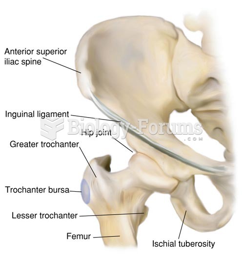 Anatomy of the Hip Joint
 Gout of the finger joint.
 Space-Beach Wallpaper
 Joint effusion of the hand
 Hubble Space telescope