A PA axial, ulnar-deviated wrist projection with poor positioning demonstrates a closed scapho-capitate joint and an open hamate-capitate joint. How should the positioning setup be adjusted for an optimal image to be obtained?
a. Increase the degree of external wrist rotation.
b. Decrease the degree of external wrist rotation.
c. Extend the fingers, with hand flat.
d. Adjust the degree of central ray angulation.
Question 2
When the patient ulnar-deviates for a PA axial, ulnar-deviated wrist projection, the
1. first metacarpal and radius are aligned.
2. distal scaphoid shifts anteriorly.
3. lunate is demonstrated distal to the radius.
4. distal scaphoid shifts posteriorly.
a. 1 and 2 only
b. 3 and 4 only
c. 1, 3, and 4 only
d. 1, 2, and 3 only

 Gunnera growing on nitrogen-poor soil.
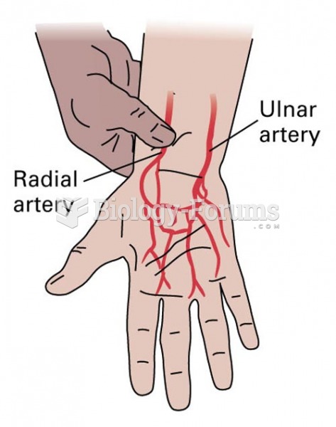 Allen Test: A patent ulnar artery reveals the return of palm perfusion despite radial artery compres
 Muscle Strength of the Wrist
 An X-ray of arthritic joints of the wrist and hand.
 Mobilize hand and wrist joints. Apply scissoring to knuckles and figure-8s to fingers. Holding the ...