|
|
|

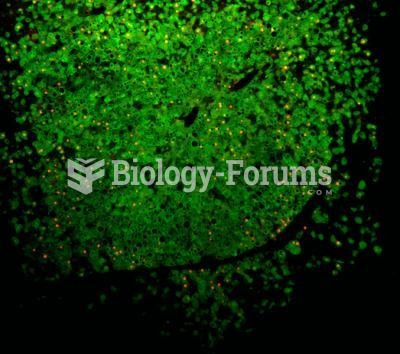 Mouse with human liver cells
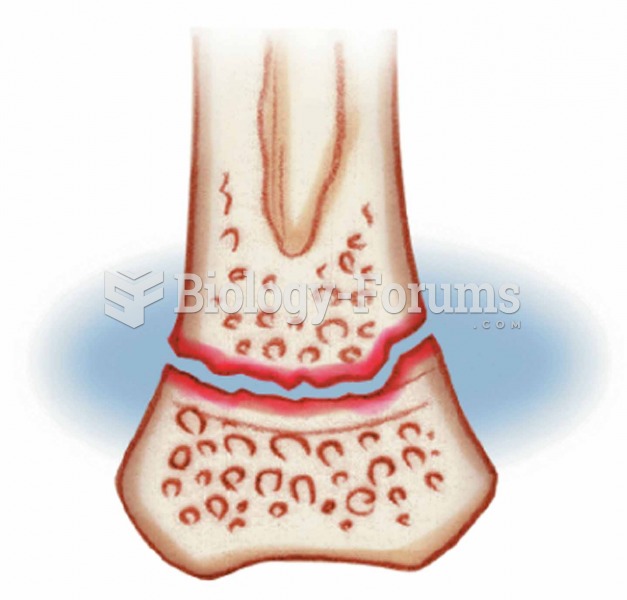 Epiphyseal Usually occurs through the growth plate where the matrix is undergoing calcification and
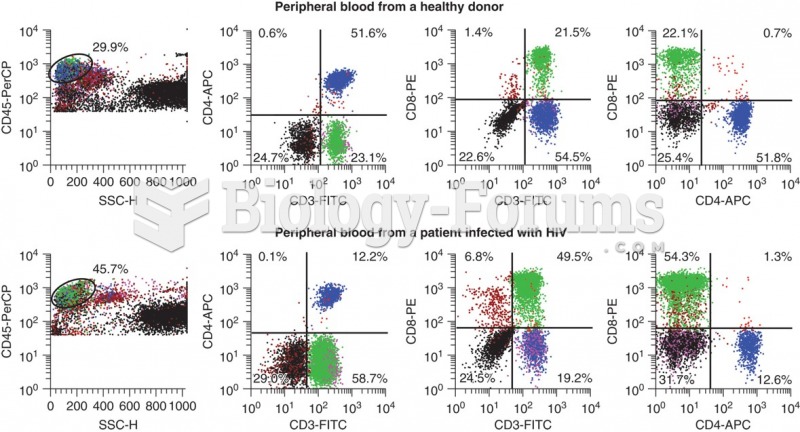 Flow cytometry data that shows the differences in CD3+ CD4+ cells that occur with HIV infection. ...
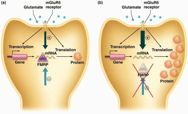 Action of FMRP protein in normal and fragile-X cells
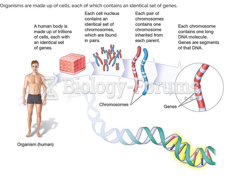 1.6 Organisms are made up of cells, each of which contains an identical set of genes.
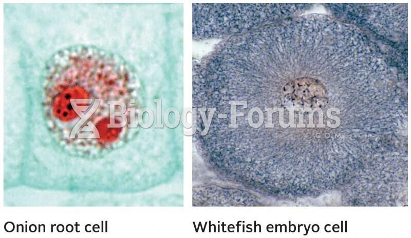 Interphase in Plant and Animal Cells