Answer to Question 1
In the first brain imaging methods, computerized axial tomography (CT), the images of the brain are collected on X-ray film. These images are then processed by a computer to generate visually and physically meaningful images. In magnetic resonance imaging (MRI) the image is derived from signals generated by changes in the levels of electromagnetic radiation from tissues under observation. Magnetic resonance spectroscopy (MRS) extends the capacity of the MRI to permit the study of tissue chemistry and metabolic function. Single photon emission computed tomography (SPECT) provides direct measurement of blood flow and several specific physiological and neurochemical features of the brain.
Answer to Question 2
b

 Differentiation of Major Headache Types
 Anterior view of major muscles.
 The major structures of the limbic system: amygdala, hippocampus, cingulate cortex, fornix, septum, ...
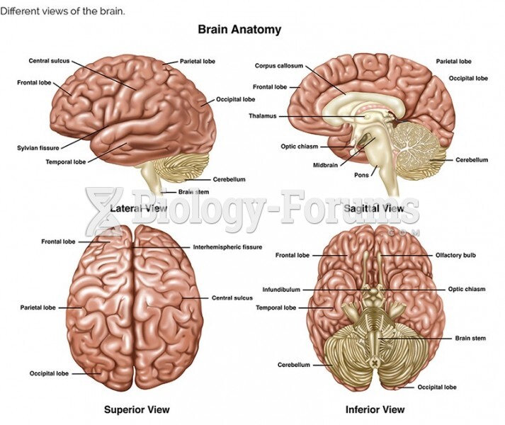 Different views of the brain