|
|
|

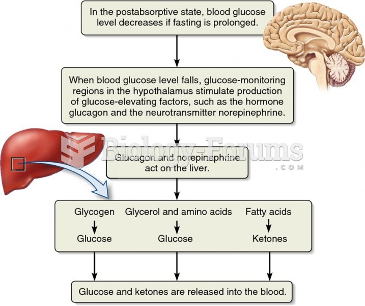 The role of the liver in fuel supply to the blood.
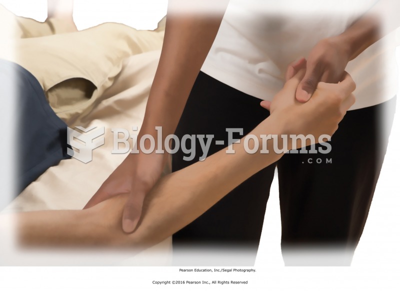 Thumb web–forearm stimulation. Using the thumb and finger of your left hand, squeeze the webbing ...
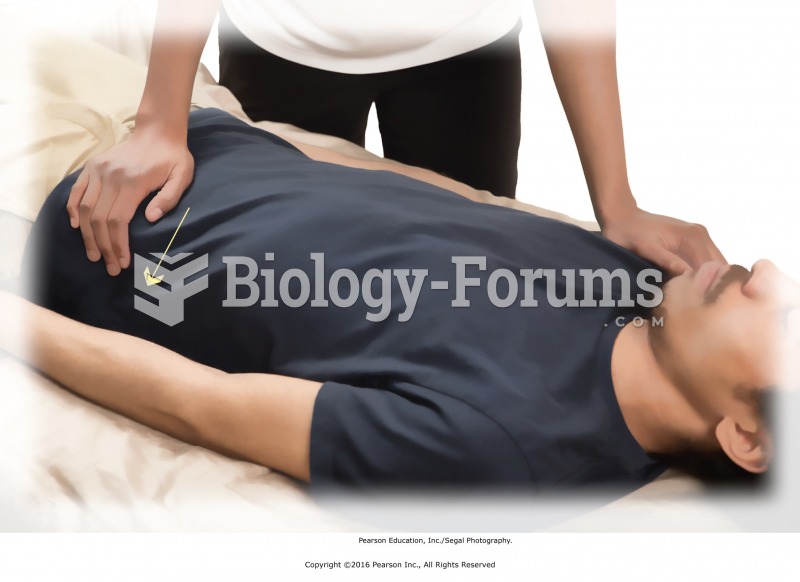 Pelvic rock. Place your right hand over the recipient’s left hip bone. Place your left hand over ...
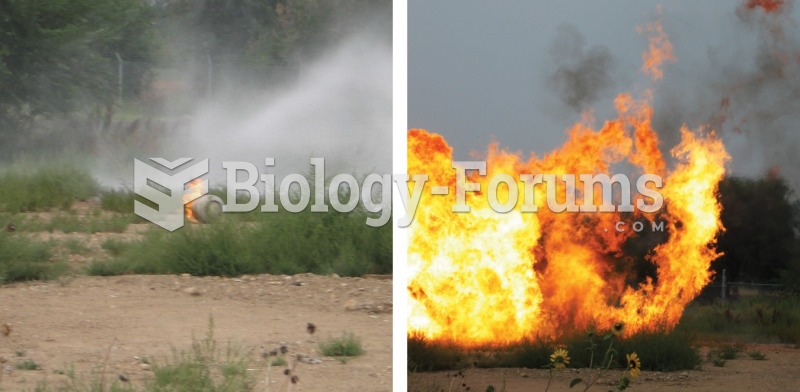 A boiling-liquid, expanding-vapor explosion (BLEVE). The burning propane cylinder on the left is not ...
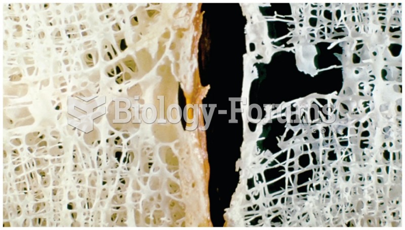 Healthy Bone (left) vs Weakened Bone (right)
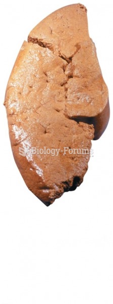 Fatty Liver