|
|
|

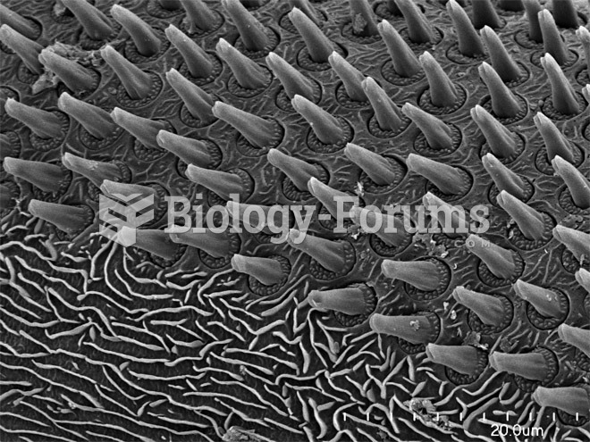 Scorpions, Spiders and Sharks: Electron-Microscope Images
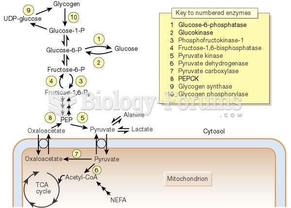 Liver Gluconeogenesis
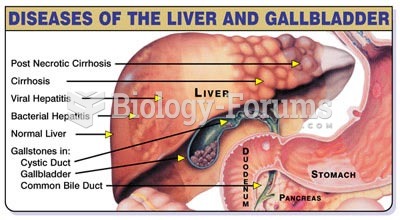 Diseases and Effects on Liver
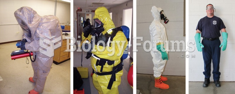 The four levels of PPE according to the EPA (from left to right): Level A, Level B, Level C, and ...
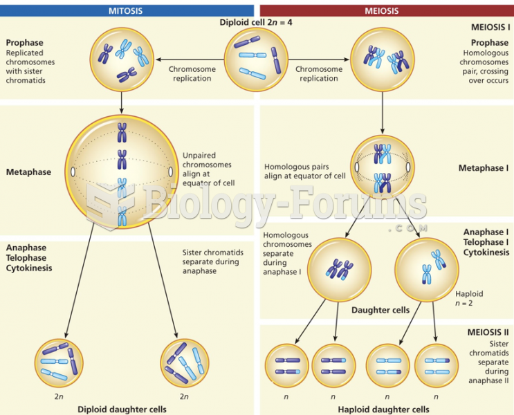 A comparison of the events in mitosis (left) and meiosis (right)
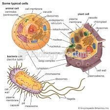 IMAGES THAT DESCRIBE CELLULAR PARTS