This topic contains a solution. Click here to go to the answer
|
|
|

 Obturator Muscle Test
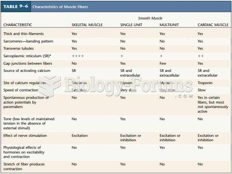 Characteristics of Muscle Fibers
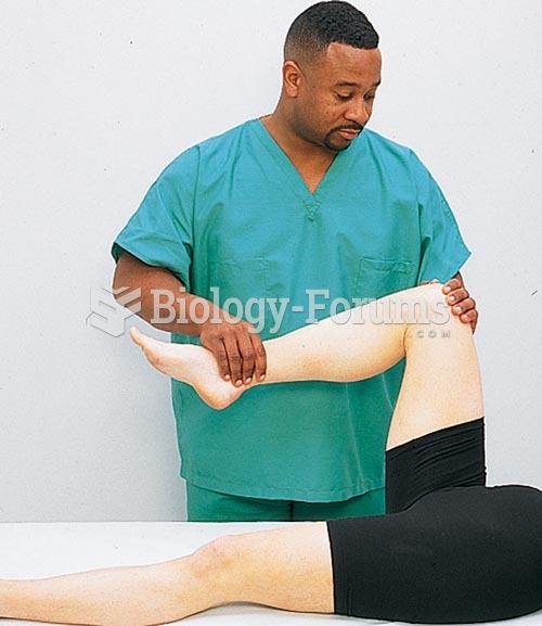
 Testing muscle strength using opposing force
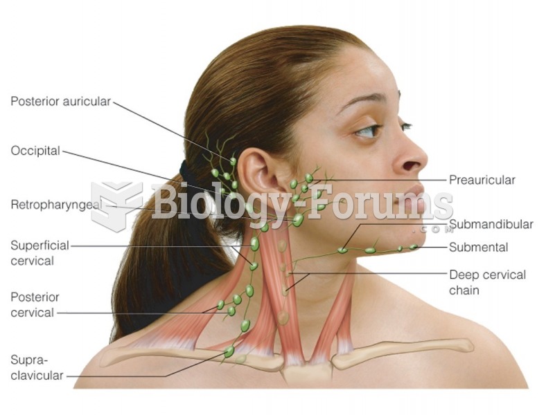 Lymph nodes of the head and neck
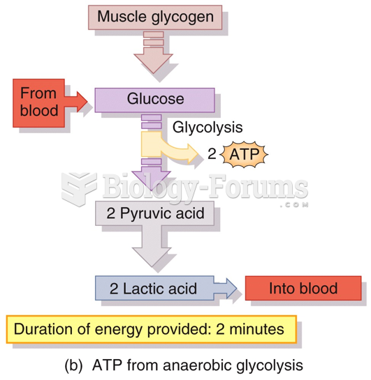 Production of ATP in Skeletal Muscle
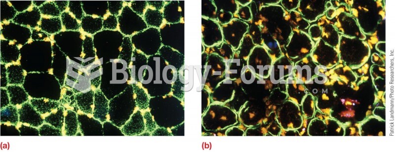 Distribution of dystrophin in muscle cells. (a) In normal muscle cells, all the dystrophin is locate