This topic contains a solution. Click here to go to the answer
|
|
|

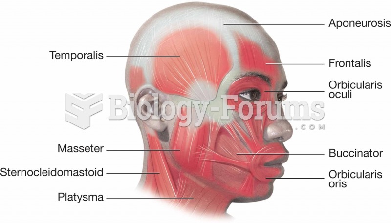 Muscles of the head and neck.
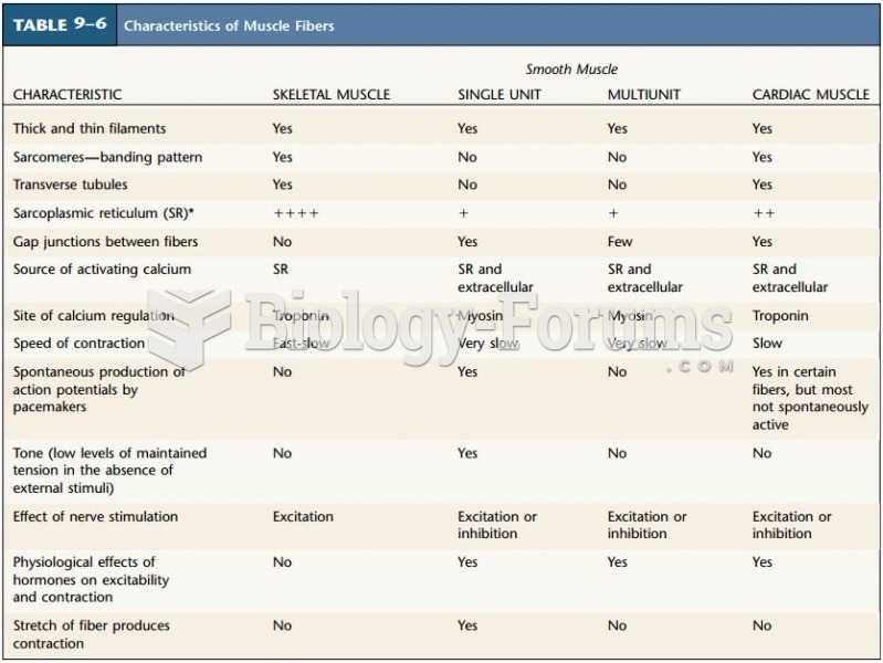 Characteristics of Muscle Fibers
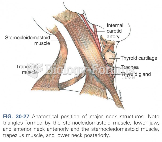 Anatomical position of major neck structures
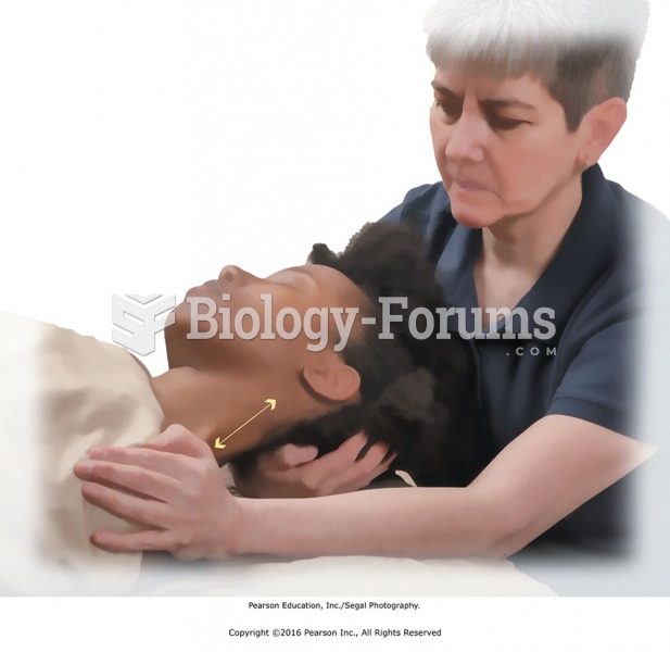 Stretch of trapezius and cervical muscles with neck in lateral flexion.
 Neck and Occipital Ridge. Apply thumb compression across the area at the base of the big toe. One ...
 Trapezius muscle movement