This topic contains a solution. Click here to go to the answer
|
|
|

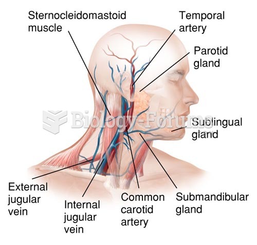 Major Veins and Arteries of the Neck
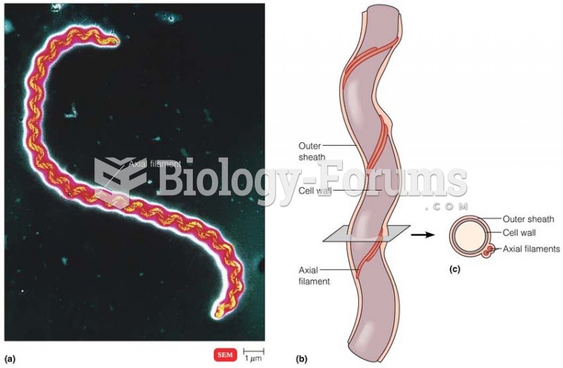 Axial Filaments
 Testing the muscle strength of the wrist
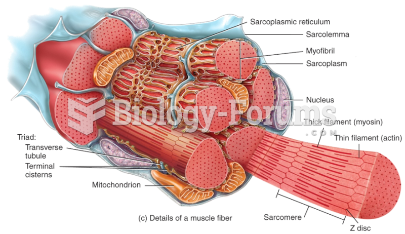 Microscopic Anatomy of a Muscle
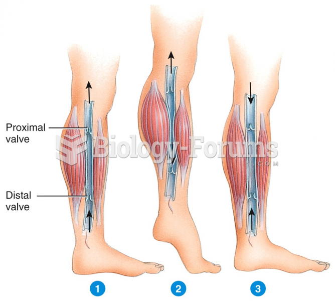 Skeletal Muscle Pump
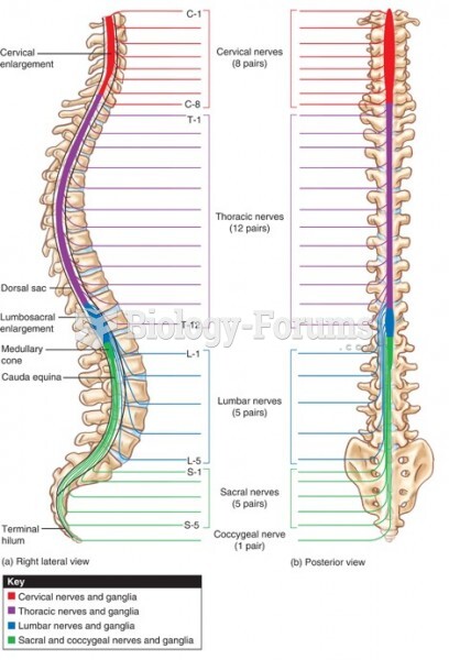 Illustration of the Spinal Nerves (a) Laterally and (b) Posteriorly. The Ganglia Are Detailed as Wel