Answer to Question 1
ANSWER:
Sound waves enter the outer ear at the pinna, which collects and focuses sounds. Sounds collected by the pinna are then channeled through the auditory canal, which ends at the tympanic membrane, or eardrum, at the boundary between the outer and middle ear. The sound waves then reach three tiny bones in the middle ear known as ossicles. The ossicles amplify sound wave input by transferring sound energy from the air of the outer and middle ear to the fluid found in the inner ear via the oval window.
The inner ear contains a fluid filled cavity known as the cochlea, which contains specialized receptor cells that respond to vibrations transmitted to the inner ear. Vibrations transmitted by the bones of the middle ear to the oval window produce waves in the fluid of the vestibular canal of the cochlea that travel around the apex and back through its tympanic canal. As waves travel through the cochlea, the basilar membrane responds with its own wavelike motion. The movement of the basilar membrane causes the hair cells of the organ of Corti to move back and forth within the fluid of the cochlear duct. Bending the hair cells stimulates the release of neurotransmitters onto the cells of the auditory nerve.
Answer to Question 2
ANSWER:
Light first passes through the cornea, which begins the process of bending light to form an image on the back of the eye. Traveling light next enters the pupil, which is actually an opening formed by the muscles of the iris. The iris adjusts the opening of the pupil in response to the amount of light present in the environment and to signals from the autonomic nervous system. Directly behind the pupil and iris is the main optical instrument of the eye, the lens. Muscles attached to the lens can change its shape, allowing us to adjust our focus to see near or distant objects. Behind the lens is the main chamber of the eye, and located on the rear surface of this chamber is the retina, a thin but complex network of neurons specialized for the processing of light.
Located in the deepest layer of the retina are the specialized receptors, the rods and cones, which transduce the light information. These rods and cones are responsible for different aspects of vision. The rods are predominantly localized to the periphery and are more sensitive to light than the cones. Rods excel at seeing in dim light, but do not provide information about color, nor do they provide clear, sharp images. In contrast cones, which are predominately found in the fovea (center of retina), function best under bright light and provide the ability to see both sharp images and color.

 Ways to prevent heat waves
 Estimated Number of New Sexually Transmitted Infections
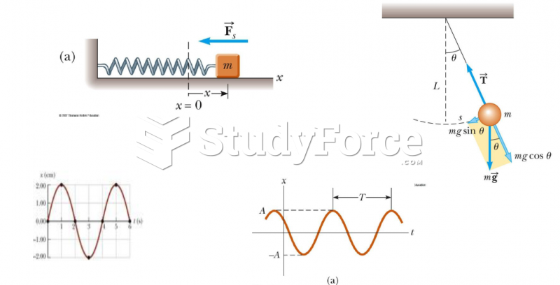 Simple harmonic motion also forms the basis for understanding mechanical waves
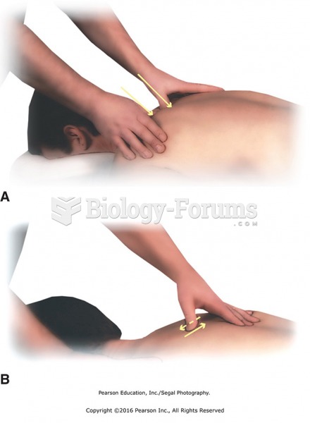 (A) Thumb presses between shoulder blades on inner and outer bladder meridian acupoints. (B) Apply ...
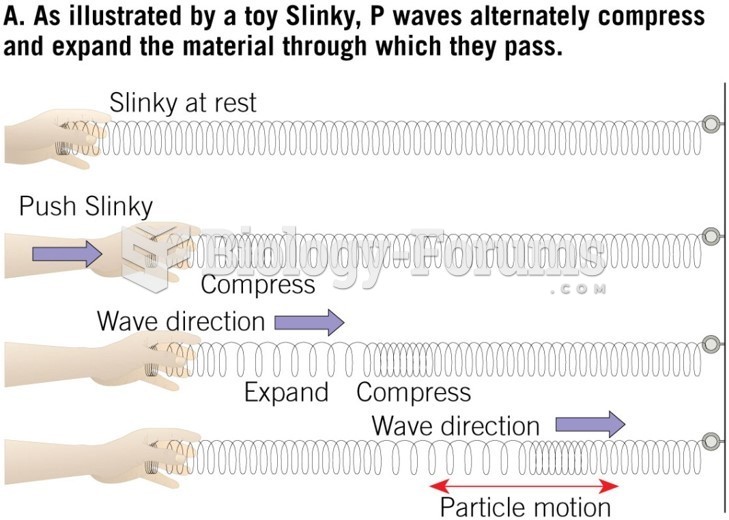 Earthquake Waves
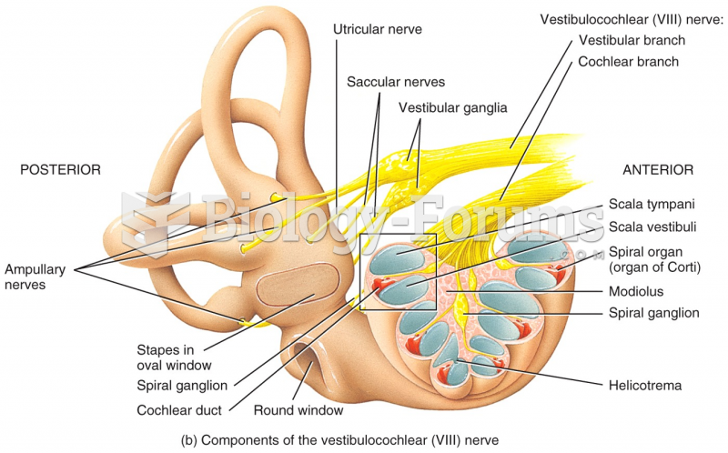 Components of the vestibulocochlear (VIII) nerve